Tibial Plateau Fractures: Anatomy, Diagnosis, and Treatment
Tibial plateau fractures are periarticular injuries of the proximal tibia frequently associated with soft tissue injury. This article provides an overview of the anatomy, epidemiology, etiology, presentation, diagnosis, treatment, potential complications, and prognosis of tibial plateau fractures:
Mohammad Hutaif
(
 ORCID ID: 0009-0001-1092-5600
) via
Google Scholar
.
ORCID ID: 0009-0001-1092-5600
) via
Google Scholar
.
__Save
__Cite
__Cited by
__Related articles
__All versions
__Import into BibTeX
Summary
Tibial plateau fractures are typically diagnosed with knee radiographs but frequently require CT scan for surgical planning. Treatment often involves ORIF in the acute setting versus delayed fixation after soft tissue swelling subsides.
Epidemiology
Incidence: Tibial plateau fractures occur in 1-2% of all fractures and have an incidence of 10.3 per 100,000 people annually. Demographics: Tibial plateau fractures occur most commonly in males in their 40s due to high-energy trauma and females in their 70s due to low-energy falls. The lateral plateau is affected in 70-80% of cases, while bicondylar and medial plateau fractures account for 10-30% and 10-20%, respectively.
Etiology Mechanism: Tibial plateau fractures are caused by the vector of applied load, amount of energy, and quality of bone. Valgus load causes lateral plateau fractures, while varus load causes medial plateau fractures. Axial load causes bicondylar fractures, and combination fractures can also occur. Fracture dislocation is associated with high-energy trauma and typically involves the medial-sided plateau. Associated conditions: Meniscal tears, ACL injuries, compartment syndrome, and neurovascular injury can all be associated with tibial plateau fractures. Soft tissue injury is frequently seen with these fractures and can impact surgical planning and outcomes.
Anatomy
Figure 1: Tibial Plateau Fractures (Source: Hutaif Orthopedic Center)
Osteology:
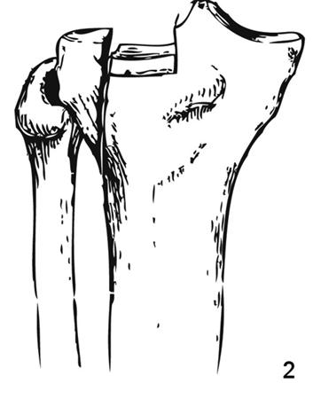
The proximal tibia is divided into the lateral and medial tibial plateau. The lateral tibial plateau is convex in shape, while the medial tibial plateau is concave. The posterior tibial slope is 6-10°, and the varus slope is 3° relative to the mechanical axis of the tibia.
Figure 2: Alignment of Proximal Tibia (Source: Hutaif Orthopedic Center)
Ligaments:
The knee joint is stabilized by several ligaments, including the ACL, PCL, MCL, and LCL. The menisci also play a role in knee stabilization.
Figure 3: Ligaments (Source: Hutaif Orthopedic Center)
Classification Schatzker classification: This classification system categorizes tibial plateau fractures into six types based on the pattern of the fracture. Type I is a lateral split fracture, Type II is a lateral split-depressed fracture, Type III is a lateral pure depression fracture, Type IV is a medial plateau fracture, Type V is a bicondylar fracture, and Type VI is a metaphyseal-diaphyseal disassociation.
Figure 4: Schatzker Classification (Source: Hutaif Orthopedic Center)
3-column concept:
This concept divides the tibial plateau into three columns: medial, lateral, and posterior. This helps to determine fixation strategy and includes posterior plateau fractures that are not considered in the Schatzker classification.
Figure 5: 3-Column Concept (Source: Hutaif Orthopedic Center)
Presentation History: The mechanism of injury, functional status, and comorbidities should be assessed. Physical exam: Examine for pain, tenderness, and swelling. Assess for any laxity on varus/valgus stress testing or instability in full extension. Evaluate for neurovascular injury and compartment syndrome.
Diagnosis and Treatment
MCQ 1
What is the first-line diagnostic tool for diagnosing tibial plateau fractures?
1. Knee radiographs
2. CT scan
3. MRI
4. Ultrasound
MCQ 2
What is the most common type of tibial plateau fracture?
1. Lateral split fracture
2. Medial plateau fracture
3. Lateral split-depressed fracture
4. Bicondylar fracture
MCQ 3
Which ligaments are primary stabilizers of the knee joint?
1. PCL and LCL
2. ACL and MCL
3. PCL and MCL
4. ACL and LCL
MCQ 4
What is the goal of treatment for tibial plateau fractures?
1. Elimination of pain
2. Restoration of range of motion
3. Restoration of alignment and congruent articular surface
4. Prevention of post-traumatic arthritis
MCQ 5
What is the most common complication associated with tibial plateau fractures?
1. Knee stiffness
2. Loss of reduction
3. Post-traumatic arthritis
4. Compartment syndrome
MCQ 6
What is the most common type of tibial plateau fracture in females?
1. Lateral split fracture
2. Medial plateau fracture
3. Lateral split-depressed fracture
4. Bicondylar fracture
Previous Next

