The shoulder girdle is a complex joint that connects the upper extremity to the axial skeleton. It is composed of four bones: the sternum, clavicle, scapula, and humerus. The shoulder girdle allows for a wide range of motion, which is essential for activities such as reaching, lifting, and throwing.
Mohammad Hutaif
,
Emial
General introduction
> 1. Complex joint
- Helps position the arm in space
- Essential in allowing us to interact with the environment
- Connects the axial skeleton to the upper extremity. Bones and joints > 1. Shoulder girdle is composed of four bones:
- Sternum
- Clavicle
- Scapula
- Humerus.
- Three major articulations: > 1. Sternoclavicular (SC) joint
- Acromioclavicular (AC) joint
- Glenohumeral (GH) joint.
- Other articulations and spaces:
- Subacromial space
- Scapulothoracic bursa. Sternum 1. Connection point of the appendicular skeleton to the axial skeleton
- Bone is composed of three parts:
- Manubrium
- Body
- Xiphoid process.
- Sternal notch is a depression between the two SC joints
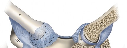
- SC joints are shallow notches at the superolateral corners of the manubrium ( ▶ Fig. 1.1)


Fig. 1.1 Diagram of the sternoclavicular (SC) joint. 1CC, first costal cartilage (ossified); 2CC, second costal cartilage; M, manubrium; 1, interclavicular ligament; 2, articular disc; 3, costoclavicular ligament (posterior lamina); 4, sternocostal joint; 5, manubriosternal joint; 6, anterior sternoclavicular ligament; 7, costoclavicular ligament (anterior lamina).- The body and manubrium serve as insertion points for the costal cartilages of ribs 1-7
- Important to understand role of SC articulation in shoulder biomechanics. Clavicle 1. Bone that spans from the sternum to the acromion
- Flat near the lateral third but becomes more convex medially
- Begins ossifying at 5 weeks in utero
- The medial epiphysis of the clavicle is the last to fuse at approximately 23-25 years of life
- The size of the bone changes in cross section at different points:
- 23 mm × 22 mm at the sternal end
- 12 mm × 12 mm at the diaphysis
- 21 mm × 11 mm at the lateral end.
- The coracoclavicular and AC ligaments stabilize the clavicle ( ▶ Fig. 1.2):
- The conoid and trapezoid ligaments provide the primary restraint in the craniocaudal direction
- The AC ligaments provide restraint in the anteroposterior direction.
- Biomechanically, the clavicle acts as a strut to support the arm for activities performed away from the body
- Serves as protection for the underlying neurovascular structures:
- Can provide mechanical advantage for the myofascial sleeve around it. Scapula 1. Triangular flat bone
- Multiple prominences
- Point of fixation for several upper extremity muscles
- Has a curved contour to articulate with the rib cage
- The spine of the scapula divides the supraspinatus and infraspinatus fossae ( ▶ Fig. 1.3)
- The coracoid process is an anterior projection and an important surgical landmark:
- Sometimes called "the lighthouse" of the shoulder
- The coracobrachialis and short head of the biceps conjoined tendon have their origin in the coracoid
- The pectoralis minor inserts on the medial aspect of the coracoid ( ▶ Fig. 1.4)
- The coracoacromial and coracoclavicular ligaments also attach to the coracoid.
- The acromion process is usually easily palpable in the subcutaneous tissue at the lateral aspect of the scapula:
- Connects the clavicle to the scapula at the AC joint
- Serves as the origin of the deltoid muscle.
Fig. 1.2 Gross anatomy of the coracoclavicular ligaments. (a) Anterior view. (b) Anterior medial view. CP, coracoid process; TL, transverse ligament; SSN, suprascapular nerve; CAL, coracoacromial ligament.

Fig. 1.3 Posterior view of a left scapula.

- The scapula widens laterally into the glenoid neck and glenoid fossa:
- Glenoid anatomy is variable but usually version will range from 9.5 degrees of anteversion to 10.5 degrees of retroversion
- The mean inclination of the glenoid is usually 4 degrees of superior tilt
- Size usually 27.8 mm by 37.5 mm in men and 23.6 mm by 32.6 mm in women. Humerus 1. Extension of the shoulder joint that allows positioning of the arm in space
- The humeral head articulates with the glenoid:
- The average radius of curvature is 24 mm in the coronal plane
- The average thickness has been reported to be 19 mm
- The average articular surface diameter is 43 mm.
Fig. 1.4 Anterior view of a left scapula.

- The greater and lesser tuberosities are the attachment points of the rotator cuff ( ▶ Fig. 1.5):
- Subscapularis attaches to the lesser tuberosity
- Supraspinatus, infraspinatus, and teres minor attach to the greater tuberosity
- The biceps groove is between the tuberosities, and can be a useful landmark during surgery.
- Retroversion of the proximal humerus is variable and can be anywhere from 10 to 5 degrees. It averages approximately 30 degrees of retroversion. Sternoclavicular joint 1. Joint between medial end of the clavicle and the superolateral aspect of the sternum
- Has been described as both a ball and socket and a saddle joint
Fig. 1.5 Posterior and anterior views of the proximal humerus demonstrating the tuberosities and the bicipital groove.

- The first costal cartilage is at the inferior aspect of the SC joint
- Only bony connection of the upper extremity to the axial skeleton
- Thickenings of the capsule serve to provide ligamentous restraint
- The posterior SC ligament serves as primary restraint for the SC joint
- The medial end of the clavicle is attached to the first rib with the costoclavicular ligament which helps restrict superior migration
- There is an articular disc in the SC joint that attaches superiorly and inferiorly.
- The SC joint moves approximately 30-35 degrees in elevation and 35 degrees in flexion/extension
- Most of the motion in the SC joint occurs in the first 90 degrees of elevation. Acromioclavicular (AC) joint 1. The AC joint is the articulation between the medial end of the acromion and the lateral end of the clavicle
- The ends of the clavicle and the acromion at the AC joint are both covered in fibrocartilage ( ▶ Fig. 1.6):
- There is also a meniscoid articular disc that covers mostly the superior portion of the joint.
- The angle of the AC joint can be variable and should be considered during surgical planning
Fig. 1.6 Left shoulder: acromial side of the AC joint. The entire capsule, detached from the clavicular side, is still attached at the acromial side, making the acromioclavicular ligaments visible. ACR,acromion, articular side; AL A/C, anterior acromioclavicular ligament; IL A/C, inferior acromioclavicular ligament; PL A/C, posterior acromioclavicular ligament; SL A/C, superior acromioclavicular ligament.

- The AC ligament provides most of the anterior and posterior stability
- The coracoclavicular ligaments provide most of the vertical stability and help maintain the relationship between the clavicle and the coracoid:
- Composed of the trapezoid (anterolateral) and conoid (posteromedial) ligaments.
Glenohumeral joint- Ball and socket or "ball on golf tee" joint that serves as the articulation between the humerus and the scapula ( ▶ Fig. 1.7)
- Allows a significant amount of mobility to help position the arm in space:
- Several dynamic and static restraints to motion
- Only a small portion of the humeral head surface contacts the glenoid at any given point.
- The glenoid fossa is surrounded by a fibrocartilaginous labrum that provides stability and deepens the articular surface


Fig. 1.7 Left shoulder, frontal view. DEL, deltoid; GLEN, glenoid; HH, humeral head;
SSP, supraspinatus.- The labrum is an attachment point for the joint capsule, glenohumeral ligaments, and long head of the biceps tendon
- Capsulolabral tears can allow for increased glenohumeral translation
- Bone loss at the glenoid can decrease the size of the articular surface and lead to increased instability
- Several named thickenings of the joint capsule provide static restraint to the glenohumeral joint ( ▶ Fig. 1.8)
- The inferior glenohumeral ligament is analogous to a hammock at the inferior aspect of the shoulder joint:
- Anterior band (AIGHL)
- Posterior band (PIGHL)
- Axillary pouch (AxIGHL)
- With the arm in abduction, further external rotation will tighten the ligament and keep the humeral head centered on the glenoid.
- The middle glenohumeral ligament (MGHL) ( ▶ Fig. 1.9) limits external rotation with the arm at the side:
- The MGHL usually originates from the upper anterior glenoid, runs deep to the subscapularis, and inserts on the lesser tuberosity
Fig. 1.8 Left cadaveric shoulder demonstrating labrum and intra-articular ligaments. GHL, glenohumeral ligament.


- There are several anatomic variants of the MGHL:
- A sublabral foramen is present in 12% of cases
- A cord-like MGHL is present in 18% of cases
- A Buford complex is found in 1-2% of cases:
- Anterior superior labrum is absent and a cord-like MGHL originates from the superior labrum.
- The superior glenohumeral ligament (SGHL) limits external rotation and inferior translation:
- Originates anterior to the biceps at the supraglenoid tubercle, but this origin can be variable.
- The posterior capsule can become pathologically thickened and limit posterior translation and adduction across the body
- The coracoacromial ligament, acromion, and coracoid all serve as static stabilizers to prevent superior migration of the humeral head
Fig. 1.9 Arthroscopic image
of the middle glenohumeral ligament (MGHL) coming off at 90 degrees angle to the subscapularis tendon. Patient is in the beach chair position (image taken from the posterior portal).

Fig. 1.10 Posterior view of the shoulder demonstrating the rotator cuff musculature. SSP, supraspinatus; SS, scapular spine; ISP, infraspinatus; Tmin, teres minor.

- The glenohumeral joint is stabilized through the midrange of motion by dynamic stabilizers:
- These consist of the rotator cuff and periscapular muscles ( ▶ Fig. 1.10 and
▶ Fig. 1.11)
- The long head of the biceps may assist in depressing the humeral head, but its role has been debated
- The rotator cuff provides compression of the humeral head into the glenoid
fossa, which further increases stability. Subacromial space 1. The space between the rotator cuff and the undersurface of the acromion and deltoid
Fig. 1.11 Anterior view of the shoulder demonstrating relationship of subscapularis to the supraspinatus. RI, rotator interval; CP, coracoid process; A/C, acromioclavicular ligaments; CAL, coracoacromial ligament; TRA, trapezoid; CON, conoid.


- Several structures can be assessed ( ▶ Fig. 1.12):
- Rotator cuff
- CA ligament
- AC joint
- Acromion
- Biceps.
- Can describe morphology of acromion as Type 1 (flat), Type II (curved), or Type III (hooked) ( ▶ Fig. 1.13). Scapulothoracic bursa 1. Several bursae that glide over the ribcage
- Two major bursae:
- Scapulothoracic bursa
- Subscapularis bursa.
- Four minor bursae are not always identifiable, and usually present only in cases of pathologic scapulothoracic articulation ( ▶ Fig. 1.14).
Fig. 1.12 View of the right subacromial space from a posterior portal in beach chair position
demonstrating a supraspinatus tear.

--- Muscles 1. There are several muscles that act around the shoulder:- It can be helpful to group into regions or think of them based on origin or insertion
- The muscles that act on the shoulder usually originate from the scapula itself, the ribs and chest wall, or the spinous processes
- See ▶ Table 1.1 for more detail. Neurovascular anatomy 1. Important to understand the anatomy of the brachial plexus
- Composed of C5-T1 nerve roots
Fig. 1.13 Enface view of the scapula demonstrating acromial morphology.

- Roots, trunks, divisions, cords, and branches ( ▶ Fig. 1.15)
- Four preclavicular branches which come from rami and trunks:
- Dorsal scapular nerve
- Long thoracic nerve
- Suprascapular nerve
- Nerve to subclavius.
- Vascular anatomy of the axillary artery branches is organized by its relationship to the pectoralis minor:
- Axillary artery is a continuation of the subclavian artery
- The subclavian artery comes off the brachiocephalic trunk on the right and
aorta on the left- It becomes the axillary artery at the lateral aspect of the first rib
- Supreme thoracic artery is the only axillary artery branch that originates medial to pectoralis minor
- Deep to the pectoralis minor, it gives off branches to the thoracoacromial
artery and the lateral thoracic arteries:- Thoracoacromial has four branches:
- Acromial
- Clavicular
- Pectoral
- Deltoid.
- Lateral to the pectoralis minor, it branches into the posterior circumflex, anterior circumflex, and subscapular arteries.
Fig. 1.14 Multiple named bursae around the shoulder, anteroposterior (AP) view.

Table 1.1 Muscle origins, insertions, and innervations about the shoulder
Muscle
| Origin
| Insertion
| Function
| Innervation
Pectoralis minor
| Ribs three to five
| Medial aspect of the coracoid
| Protracts scapula
| Medial pectoral
nerve
Pectoralis major
| Sternum, clavicle,
ribs
| Lateral aspect of the bicipital groove
| Adducts and internally rotates arm
| Medial and lateral
pectoral nerves
Latissimus dorsi
| Spinous processes of thoracolumbar spine and ilium
| Medial aspect of the bicipital groove
| Extends, adducts, internally rotates humerus
| Dorsal scapular nerve
Trapezius
| Spinous processes
C7-C12
| Posterior surface lateral third clavicle, acromion, scapular spine
| Rotates scapula
| Spinal accessory nerve (cranial nerve XI)
Rhomboid
major
| Spinous processes
T2-T5
| Medial border
scapula
| Adducts
scapula
| Dorsal scapular nerve
Rhomboid
minor
| Spinous processes
C7-T1
| Medial aspect of
scapular spine
| Adducts
scapula
| Dorsal scapular nerve
Levator scapulae
| Transverse processes C1-C4
| Superomedial
scapula
| Move scapula cephalad and rotate
| C3 and C4
Subclavius
| First rib
| Inferior clavicle
| Moves clavicle caudally
| Upper trunk brachial
plexus
Serratus anterior
| First through
ninth rib
| Ventral medial surface of scapula
| Holds scapula against chest wall
| Long thoracic nerve
Deltoid
| Lateral aspect clavicle, acromion, and scapular spine
| Deltoid tuberosity of humerus
| Abducts arm
| Axillary nerve
Teres major
| Inferior scapula
| Medial aspect bicipital groove
| Adducts, internally rotates, extends arm
| Lower subscapular
nerve
Subscapularis
| Ventral scapula
| Lesser tuberosity
| Internally
rotates arm
| Upper and lower subscapular nerves
Supraspinatus
| Supraspinous fossa
| Greater
tuberosity
| Abducts and internally rotates arm
| Suprascapular nerve
Infraspinatus
| Infraspinous fossa
| Greater
tuberosity
| Externally rotates arm
| Suprascapular nerve
Teres minor
| Dorsolateral scapula
| Greater
tuberosity
| Externally rotates arm
| Axillary nerve
Biceps brachii
| Supraglenoid
| Radial
| Flex and supi-
| Musculocutaneous
| tubercle (long
| tuberosity
| nate elbow
| nerve
| head) and cora-
| |
| |
coid (short head)
| |
| |


Fig. 1.15 Diagram of the brachial plexus. Suggested Readings
Hoppenfeld S, de Boer P, Buckley R. The shoulder. In: Surgical Exposures in Orthopaedics: The Anatomic Approach. Philadelphia, PA: Lippincott Williams & amp; Wilkins; 20
