Shoulder Dislocation and Greater Tuberosity Fracture Diagnosis and Management
2. How would you treat and manage this condition? Show Answer Show Explanation
3. What are the risks and complications you anticipate? Show Answer Show Explanation
4. What would you do if attempted closed reduction fails and the patient is in extended pain? Show Answer Show Explanation
5. How will you perform the shoulder reduction? Show Answer Show Explanation
6. Other than the described risk factors, what factors can also prevent closed stable reduction of the dislocation? Show Answer Show Explanation
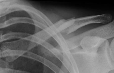
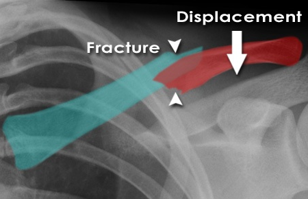
7. The X-ray of the right shoulder, one week later, displays no humeral neck fracture but significant greater tuberosity displacement. What is your management strategy? Show Answer Show Explanation
8. Highlight the risks of non-operative management of displaced greater tuberosity fracture. Show Answer Show Explanation
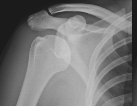
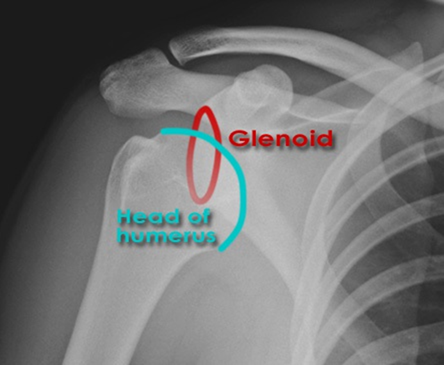
Anterior shoulder dislocation - AP view
Humeral head and glenoid surfaces are not aligned The humeral head lies below the coracoid
Anterior shoulder dislocation - Y view
The humeral head lies anterior to the glenoid and inferior to the coracoid process
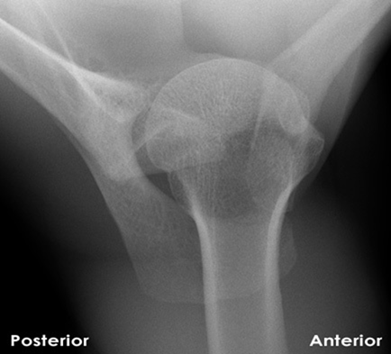
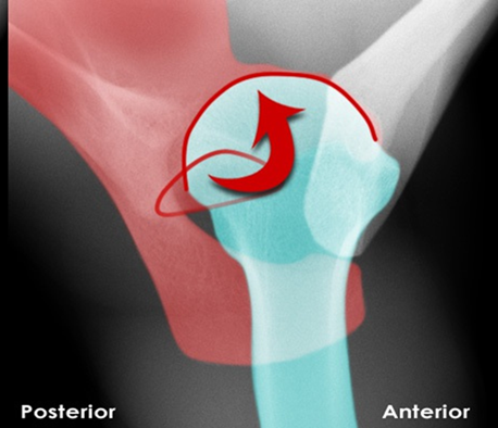
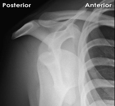
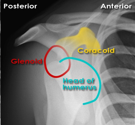
Anterior shoulder dislocation - Axial view
The humeral head surface is no longer aligned with the glenoid The humeral head lies anterior to the glenoid
Anterior shoulder dislocation - AP view
Humeral head and glenoid surfaces are not aligned
The humeral head lies below the coracoid
Anterior shoulder dislocation - Y view
The humeral head lies anterior to the glenoid and inferior to the coracoid process
Anterior shoulder dislocation - Axial view
The humeral head surface is no longer aligned with the glenoid The humeral head lies anterior to the glenoid
