Comprehensive Introduction and Patho-Epidemiology
Intractable plantar keratosis (IPK) represents a localized, severely painful hyperkeratotic lesion on the plantar aspect of the foot, most commonly located beneath one or more of the lesser metatarsal heads. Unlike diffuse callosities caused by generalized friction or inappropriate footwear, an IPK is the direct clinical manifestation of altered forefoot biomechanics—specifically, a focal concentration of weight-bearing forces during the stance and propulsive phases of the human gait cycle. The pathology is fundamentally mechanical, driven by a structural abnormality within the forefoot that creates an unyielding focal point of pressure against the plantar skin during the repetitive loading of ambulation.
The primary structural etiology for a discrete, nucleated IPK is an abnormally long or rigidly plantarflexed lesser metatarsal. Under physiologic conditions, the metatarsal heads share the load of the forefoot in a harmonious parabolic curve, often referred to as the Maestro curve. When a single metatarsal projects distally beyond this harmonious curve, or when it is fixed in a plantarflexed posture relative to its neighbors, it acts as a rigid, unyielding pivot point. Consequently, this offending metatarsal head absorbs a disproportionate quantum of ground reaction force (GRF) during terminal stance and pre-swing phases. Over time, this chronic mechanical overload induces a protective, albeit pathologic, hypertrophy of the stratum corneum.
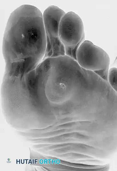
Clinical presentation of a severe, nucleated intractable plantar keratosis located beneath the second metatarsal head.
This hypertrophic response leads to the formation of a dense, nucleated keratinous plug that drives deeply into the underlying dermis. The invagination of this hard keratin mass compresses the highly innervated dermal tissue, causing debilitating, sharp, and localized pain that patients often describe as "walking on a stone." Epidemiologically, IPKs are most frequently observed beneath the second metatarsal head, particularly in patients with a Morton's foot structure (where the second metatarsal is significantly longer than the first) or in the setting of hallux valgus, where the first ray becomes biomechanically incompetent, leading to transfer metatarsalgia to the lesser rays.
When conservative measures—such as custom orthoses with metatarsal offloading pads, aggressive scalpel debridement of the hyperkeratotic nucleus, and footwear modifications including rigid rocker-bottom soles—fail to provide durable relief, surgical intervention becomes indicated. The Shortening Oblique Metatarsal Osteotomy, originally described by Giannestras and subsequently refined by Mann, is a powerful, structurally corrective procedure. It is designed to definitively decompress the affected metatarsophalangeal (MTP) joint by shortening the metatarsal shaft, thereby restoring a physiologic load distribution across the forefoot and eliminating the mechanical genesis of the keratosis.
Detailed Surgical Anatomy and Biomechanics
A profound understanding of forefoot anatomy and biomechanics is an absolute prerequisite for executing a successful metatarsal osteotomy. The lesser metatarsals are long, tubular bones consisting of a base, diaphysis, metaphysis, and head. They articulate proximally with the cuneiforms and cuboid at the tarsometatarsal (TMT) joints, and distally with the proximal phalanges at the MTP joints. The second metatarsal is uniquely keyed into the mortise created by the medial, intermediate, and lateral cuneiforms, rendering its TMT joint exceptionally rigid. This inherent rigidity is a primary reason why the second metatarsal is highly susceptible to mechanical overload; it lacks the sagittal plane compliance seen in the first, fourth, and fifth rays.
The vascular anatomy of the lesser metatarsals is of paramount importance to the orthopedic surgeon, particularly when performing diaphyseal osteotomies. The blood supply is derived from two primary sources: the nutrient artery and the periosteal plexus. The principal nutrient artery typically enters the lateral aspect of the middle third of the metatarsal diaphysis, supplying the endosteal circulation. The periosteal plexus is fed by branches of the dorsal and plantar metatarsal arteries. Because the shortening oblique osteotomy is performed in the diaphyseal region, it inherently disrupts the medullary blood supply. Consequently, the healing of the osteotomy becomes critically dependent on the integrity of the periosteal plexus. Excessive subperiosteal stripping during the surgical approach can devascularize the bone fragments, drastically increasing the risk of delayed union, nonunion, or avascular necrosis of the capital fragment.
Biomechanically, the metatarsal heads function as the primary weight-bearing fulcrum of the forefoot during the propulsive phase of gait. The windlass mechanism, driven by the plantar fascia, stabilizes the longitudinal arch and tightly packs the metatarsal heads against the ground. If a metatarsal is excessively long, it disrupts the transverse metatarsal arch. The ground reaction forces, which should ideally be distributed across the metatarsal heads in a 2:1:1:1:1 ratio (with the first metatarsal bearing twice the load of each lesser metatarsal), are pathologically concentrated. Furthermore, the intrinsic musculature, including the lumbricals and interossei, rely on precise metatarsal length for optimal tensioning. Altering this length through osteotomy not only decompresses the plantar skin but also subtly alters the tension in these intrinsic muscles, which must be accounted for during preoperative planning to prevent secondary toe deformities.
Exhaustive Indications and Contraindications
The decision to proceed with a shortening oblique metatarsal osteotomy must be based on a rigorous evaluation of the patient's pathology, failed conservative management, and overall physiological status. The primary indication is a recalcitrant, nucleated intractable plantar keratosis located directly beneath a lesser metatarsal head, driven by an anatomically long metatarsal. The surgeon must confirm that the pain is isolated to the IPK and that the keratosis is the direct result of a structural length discrepancy rather than a systemic dermatological condition, a rigid plantarflexed deformity, or generalized fat pad atrophy.
Conservative management must be exhausted prior to surgical consideration. This typically involves a minimum of six months of non-operative care, including regular podiatric debridement, the utilization of custom-molded orthoses featuring specific metatarsal offloading accommodations (such as a U-shaped pad or metatarsal bar), and the modification of footwear to include stiff-soled shoes with a rocker bottom to decrease forefoot pressures during the terminal stance phase of gait. Only when these interventions fail to provide acceptable symptomatic relief, and the patient's quality of life remains significantly impaired, should surgical shortening be entertained.
Contraindications must be meticulously respected to prevent catastrophic postoperative complications. Absolute contraindications include active localized or systemic infection, severe peripheral arterial disease (PAD) compromising the healing potential of the distal extremity, and profound peripheral neuropathy (such as advanced diabetic neuropathy), which negates the patient's ability to provide protective feedback and dramatically increases the risk of Charcot neuroarthropathy or unrecognized hardware failure. Relative contraindications include heavy tobacco use, osteopenia or osteoporosis which may compromise hardware purchase, and psychiatric conditions that preclude adherence to strict postoperative non-weight-bearing or protected weight-bearing protocols.
| Category | Specific Condition/Factor | Clinical Rationale |
|---|---|---|
| Absolute Indications | Recalcitrant IPK beneath a long lesser metatarsal | Mechanical overload due to length discrepancy requires structural shortening for definitive resolution. |
| Absolute Indications | Failure of >6 months of conservative therapy | Ensures surgery is reserved for truly intractable cases unresponsive to orthoses and debridement. |
| Absolute Contraindications | Active deep space infection or osteomyelitis | Introduction of hardware in an infected field guarantees surgical failure and potential amputation. |
| Absolute Contraindications | Severe Peripheral Arterial Disease (PAD) | Inadequate perfusion will result in wound dehiscence, nonunion, and potentially gangrene. |
| Absolute Contraindications | Profound sensory neuropathy | Lack of protective sensation leads to unrecognized hardware failure, nonunion, and Charcot arthropathy. |
| Relative Contraindications | Active, heavy tobacco use | Nicotine causes profound microvascular vasoconstriction, exponentially increasing the risk of diaphyseal nonunion. |
| Relative Contraindications | Severe osteoporosis | Poor bone stock compromises the purchase of interfragmentary lag screws, risking malunion. |
| Relative Contraindications | Generalized plantar fat pad atrophy | Shortening one metatarsal in the setting of global atrophy will predictably cause transfer metatarsalgia. |
Pre-Operative Planning, Templating, and Patient Positioning
Clinical Assessment
A meticulous clinical examination is paramount and serves as the foundation of preoperative planning. The orthopedic surgeon must first definitively differentiate an IPK from other plantar lesions, most notably a plantar wart (verruca plantaris) or an epidermal inclusion cyst. An IPK will typically present directly beneath a bony prominence, exhibit maximum tenderness with direct vertical pressure (rather than the side-to-side pinching pain characteristic of a verruca), and reveal a deep, avascular central keratinous nucleus upon paring with a scalpel. In contrast, a verruca will often display punctate bleeding (thrombosed capillaries) when pared.
The surgeon must thoroughly evaluate the mobility of the tarsometatarsal (TMT) joints and the metatarsophalangeal (MTP) joints. The sagittal plane mobility of the offending ray is critical. A rigid, plantarflexed metatarsal with a normal length profile requires a fundamentally different surgical approach—such as a basilar dorsiflexory wedge osteotomy—compared to a metatarsal that is simply excessively long but normally aligned in the sagittal plane. Furthermore, the presence of concurrent deformities, such as hallux valgus or hammertoes, must be documented, as addressing an isolated lesser metatarsal without correcting a globally incompetent first ray will inevitably lead to recurrent transfer lesions.
Radiographic Evaluation
Standard weight-bearing radiographs of the foot—comprising anteroposterior (AP), lateral, and axial sesamoid/forefoot views—are mandatory for accurate preoperative templating. Non-weight-bearing films are virtually useless for evaluating forefoot biomechanics, as they fail to demonstrate the dynamic splaying and functional length of the metatarsals under load.
- Anteroposterior (AP) View: This view is utilized to assess the relative lengths of the metatarsals. The normal metatarsal parabola dictates that the second metatarsal is slightly longer than the first, with a smooth, cascading decrease in length from the second to the fifth metatarsal. An abrupt discrepancy, where a lesser metatarsal protrudes distally beyond this cascade, indicates the offending digit. The surgeon must measure the exact length discrepancy to determine the required shortening.
- Lateral View: The lateral projection is essential to evaluate the sagittal alignment of the metatarsals, ruling out structural plantarflexion at the TMT joint or dorsal subluxation at the MTP joint.
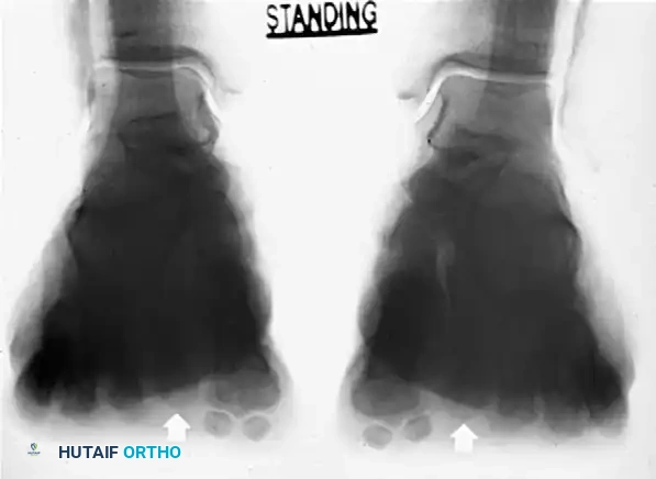
- Axial Forefoot View: This specialized view is critical for visualizing the coronal plane alignment of the metatarsal heads. It allows the surgeon to identify any abnormal plantar prominence of a specific metatarsal head relative to its neighbors, directly correlating the radiographic anatomy with the clinical location of the IPK.
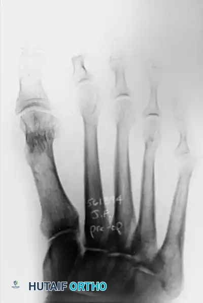
Preoperative weight-bearing AP radiograph demonstrating an excessively long second metatarsal, disrupting the normal forefoot parabola.
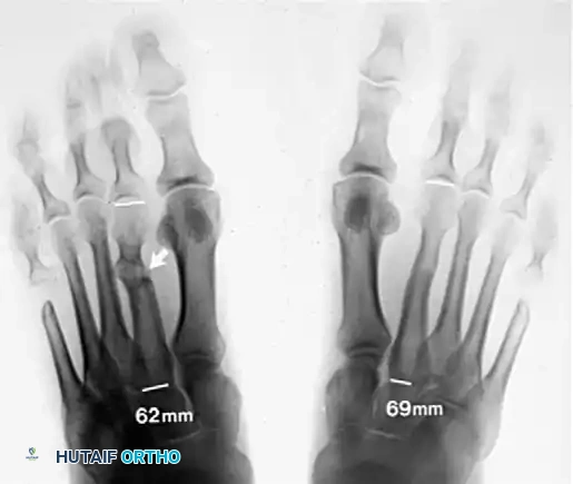
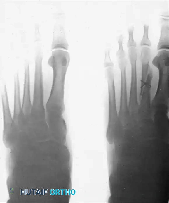
Comparative radiographic evaluation highlighting the length discrepancy. Precise measurement (e.g., 69mm vs. 62mm) is critical for determining the exact amount of shortening required.
Axial standing forefoot radiographs. Note the abnormal plantar projection and elevation dynamics of the metatarsal heads (arrows), which correlate directly with the location of the plantar keratosis.
Preoperative templating should determine the exact amount of shortening needed—usually not more than 5 to 6 mm. Over-shortening is a catastrophic and highly predictable error that leads to transfer metatarsalgia beneath the adjacent, now relatively longer, metatarsal heads.
Patient Positioning and Anesthesia
The procedure is typically performed on an outpatient basis. Anesthesia options include a regional block (such as an ankle block or a popliteal fossa block) supplemented with intravenous sedation, or general anesthesia, depending on patient preference and comorbidities. The patient is positioned supine on the operating table. A well-padded calf or thigh tourniquet is applied to ensure a bloodless surgical field, which is critical for identifying delicate neurovascular structures and confirming precise osteotomy reduction. Following the administration of prophylactic intravenous antibiotics, the entire foot and ankle are subjected to standard orthopedic preparation and draping.
Step-by-Step Surgical Approach and Fixation Technique
The modified Giannestras/Mann technique utilizes a diaphyseal oblique osteotomy. The oblique nature of the cut is biomechanically superior to a simple transverse osteotomy for several reasons: it provides a significantly larger surface area for endosteal and periosteal bone healing, it inherently resists dorsal displacement of the capital fragment during weight-bearing, and it allows for the application of rigid interfragmentary lag screw compression, which drastically reduces the risk of nonunion.
Surgical Approach and Dissection
A 5-cm longitudinal dorsal incision is made, centered precisely over the diaphysis of the affected metatarsal. The incision should begin just distal to the tarsometatarsal (TMT) joint and extend distally toward the metatarsal neck. As the incision is deepened through the subcutaneous tissue, the surgeon must be hyper-vigilant in identifying and meticulously retracting the dorsal cutaneous nerve branches (typically terminal branches of the superficial peroneal nerve). Iatrogenic injury to these nerves results in painful neuroma formation, which can be more debilitating than the original IPK.
The extensor digitorum longus (EDL) and extensor digitorum brevis (EDB) tendons are identified and retracted either laterally or medially to expose the dorsal periosteum of the metatarsal. A sharp, longitudinal incision is made directly through the periosteum. Using a Freer or Key elevator, a subperiosteal dissection is performed circumferentially around the shaft of the metatarsal. The surgeon must strictly limit the subperiosteal stripping to the diaphyseal region required for the osteotomy and subsequent fixation. Excessive stripping, particularly distal extension toward the metatarsal neck, severely compromises the vascular supply to the capital fragment and exponentially increases the risk of delayed union or avascular necrosis.
The "Two Nicks" Measurement Technique
Accurate shortening is the single most critical step of this procedure. Once the bone is cut, the oblique angle and the bone removed by the saw blade (the saw kerf) make it exceptionally difficult to visually judge how much length has been resected. To eliminate this guesswork, the "Two Nicks" technique is employed. Prior to making the osteotomy, a microsaw or a sharp osteotome is used to make two small, transverse cortical "nicks" on the dorsal aspect of the metatarsal shaft. These nicks must be placed at a precise, fixed distance from each other (e.g., exactly 10 mm apart), spanning the planned osteotomy site. After the osteotomy is performed and the bone is shortened, the distance between these two nicks is measured again using a sterile caliper. If the initial distance was 10 mm, and the preoperative template dictated 5 mm of shortening, the bone fragments are manipulated until the nicks are exactly 5 mm apart.
Executing the Osteotomy
The osteotomy is executed in an oblique fashion, typically running from dorsal-proximal to plantar-distal. This specific orientation is chosen because it creates a mechanical block that resists dorsal displacement of the distal fragment when ground reaction forces are applied to the plantar aspect of the foot. The cut is performed using a small sagittal saw under continuous, copious sterile saline irrigation. Irrigation is mandatory to prevent thermal necrosis of the cortical bone, which is a leading cause of diaphyseal nonunion. The pre-calculated amount of bone (usually 5 to 6 mm) is resected as a parallel-sided wafer to ensure the distal fragment translates proximally without altering the sagittal or coronal alignment of the metatarsal head.
Reduction and Fixation
Once the bone is shortened, the fragments must be rigidly stabilized. The osteotomy is reduced and held in its newly shortened, anatomically aligned position using a small pointed bone-holding forceps (e.g., a Weber clamp). The shortening is verified by measuring the distance between the pre-marked dorsal nicks.
Fixation Option A (Wire Fixation): While largely historical, this technique remains a viable salvage option if bone quality is exceptionally poor. A transverse hole is drilled through both cortical fragments using a 0.045-inch Kirschner wire. A 20-gauge stainless steel cerclage wire is passed through the hole and wrapped around the osteotomy in a figure-of-eight fashion to secure the fixation. The sharp ends of the wire are folded flat against the metatarsal shaft to prevent soft tissue irritation.
Postoperative AP radiograph demonstrating fixation of the oblique osteotomy using the 20-gauge wire technique.
Fixation Option B (Interfragmentary Compression Screw): This is the preferred, modern gold-standard technique due to its superior biomechanical stability. A 2.0-mm or 2.4-mm cortical lag screw is placed across the osteotomy, oriented perpendicular to the cut surface to maximize compression and minimize shear forces. The near cortex is overdrilled to create a gliding hole, the far cortex is drilled with the core diameter drill bit, and the near cortex is carefully countersunk to prevent the screw head from creating a stress riser. The tract is measured, tapped, and the screw is inserted to achieve rigid interfragmentary compression.
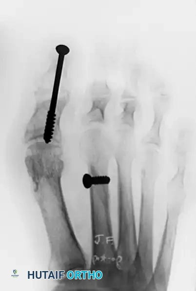
Postoperative AP radiograph showing rigid fixation of the shortened second metatarsal with an interfragmentary compression screw. Note the concurrent arthrodesis of the interphalangeal joint of the hallux for a varus deformity.
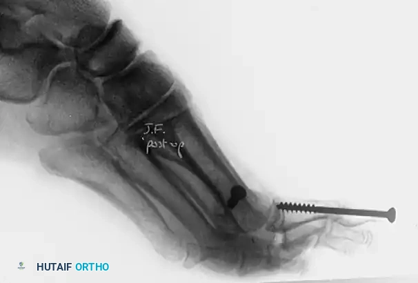
Lateral postoperative radiograph of the same patient, confirming excellent sagittal alignment of the metatarsal osteotomy and flush seating of the compression screw.
Alternative Technique: Basilar Dorsal Closing Wedge Osteotomy
In cases where the primary pathology is a rigidly plantarflexed metatarsal rather than an excessively long one, a diaphyseal shortening osteotomy is inappropriate. Instead, a basilar dorsal closing wedge osteotomy is indicated. This osteotomy is performed proximally, at the metaphyseal-diaphyseal junction. The surgeon must be exceedingly cautious not to remove more than a 2 to 3-mm dorsal wedge of bone. Removing a larger wedge will result in excessive dorsal elevation of the metatarsal head, completely offloading it and guaranteeing severe transfer metatarsalgia. Rigid internal fixation, typically utilizing a dorsal low-profile locking plate or crossed K-wires, is required.
Complications, Incidence Rates, and Salvage Management
While highly effective when executed flawlessly, metatarsal osteotomies carry a distinct and unforgiving risk profile. The diaphysis of the metatarsal has a relatively tenuous blood supply compared to the highly vascularized metaphyseal regions, making it intrinsically susceptible to healing complications. The surgeon must be prepared to identify and manage these complications aggressively.
Nonunion and delayed union are the most dreaded complications of the diaphyseal osteotomy. They are usually the direct result of technical errors: inadequate rigid fixation allowing micromotion, excessive periosteal stripping devascularizing the bone, or thermal necrosis during the saw cut due to insufficient irrigation. Patients typically present with persistent, deep dorsal midfoot pain, localized swelling, and a palpable, tender mass at the osteotomy site indicative of hypertrophic fibrous tissue.
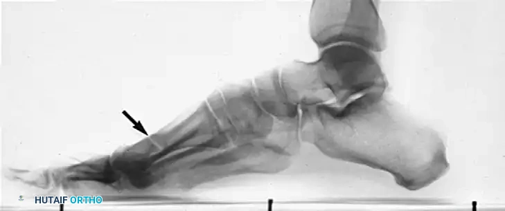
Lateral radiograph demonstrating a painful nonunion of a transverse metatarsal osteotomy (arrow). Note the dorsal angulation and hypertrophic callus formation attempting to bridge the unstable gap.
Management of a symptomatic nonunion requires definitive revision surgery. The fibrous nonunion site must be aggressively debrided down to bleeding, viable cortical bone (the "paprika sign"). Rigid internal fixation, usually in the form of a dorsal spanning plate, is applied. This must be supplemented by an autologous onlay bone graft—often harvested from the ipsilateral calcaneus or proximal tibia—to provide the necessary osteoinductive and osteoconductive properties to stimulate osteogenesis across the defect.
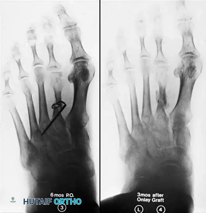
Radiographic progression of nonunion management. (Left) AP view at 6 months post-op showing failed wire fixation and nonunion. (Right) AP view 3 months after revision surgery utilizing an onlay bone graft, demonstrating successful consolidation. The patient was walking comfortably in a firm, solid shoe.
Malunion and transfer metatarsalgia represent the other major category of complications. If the metatarsal is shortened excessively (greater than 6 mm) or if the distal fragment is allowed to elevate dorsally during the healing phase due to loss of fixation, the affected metatarsal head will no longer participate in the weight-bearing triad of the forefoot. The ground reaction forces will immediately and predictably transfer to the adjacent, relatively longer metatarsal heads. This results in the rapid development of a new, often more painful IPK beneath the adjacent metatarsal. Prevention is the only acceptable management strategy, relying heavily on the "Two Nicks" technique. If severe symptomatic transfer metatarsalgia occurs, corrective osteotomy of the newly overloaded metatarsal may be required.
| Complication | Estimated Incidence | Primary Etiology | Salvage Management Strategy |
|---|---|---|---|
| Nonunion / Delayed Union | 3% - 10% | Thermal necrosis, excessive stripping, unstable fixation. | Revision debridement, rigid dorsal plating, autologous bone grafting. |
| Transfer Metatarsalgia | 5% - 15% | Over-shortening (>6mm), dorsal elevation of capital fragment. | Conservative offloading first; revision osteotomy of newly overloaded ray if refractory. |
| Hardware Irritation | 10% - 20% | Prominent screw head, superficial wire placement. | Hardware removal after complete radiographic consolidation (minimum 6 months). |
| Avascular Necrosis (AVN) | < 2% | Aggressive distal periosteal stripping devascularizing the head. | Prolonged offloading; rarely requires metatarsal head resection or arthroplasty. |
| Infection (Superficial/Deep) | 1% - 3% | Wound contamination, poor soft tissue envelope. | Oral/IV antibiotics; deep infections require immediate hardware removal and debridement. |
Phased Post-Operative Rehabilitation Protocols
Successful healing of a diaphyseal metatarsal osteotomy requires strict, unwavering adherence to postoperative immobilization protocols. The rehabilitation is phased to respect the biology of bone healing while preventing secondary complications such as joint contractures or deep vein thrombosis.
Phase 1: Immediate Post-Op (Weeks 0-4)
Immediately following surgery, the foot is placed in a bulky, sterile compressive dressing. Within the first few days, the patient is transitioned into a well-padded, short-leg cast equipped with a toe plate. The toe plate is a critical component; it extends beneath the digits to support them and prevent dorsal contracture of the MTP joints, which can occur due to the relative lengthening of the extensor tendons when the metatarsal is shortened. For the standard shortening oblique osteotomy secured with rigid interfragmentary screw fixation, protected weight-bearing in the cast (heel-touch or flat-foot walking) is generally permitted immediately. However, if wire fixation was utilized, or if intraoperative assessment revealed poor bone quality, a strict non-weight-bearing protocol is mandated for the first four weeks.
Phase 2: Transition and Early Mobilization (Weeks 4-6)
At the 4-week mark, the cast is removed. The surgical site is evaluated for clinical healing, defined as the absence of pain or micromotion upon manual palpation of the osteotomy site. Radiographs are obtained to assess for early callus formation and maintenance of hardware position. If clinical and radiographic parameters are satisfactory, the patient is transitioned to a stiff-soled postoperative shoe or a controlled ankle motion (CAM) boot. Active and passive range of motion exercises for the MTP and interphalangeal joints are initiated to combat stiffness. Weight-bearing is advanced to tolerance within the protective boot.
Phase 3: Long-Term Maturation (Weeks 6-12+)
By 6 to 8 weeks, most patients demonstrate sufficient radiographic consolidation to transition out of the CAM boot and into a standard, supportive, wide-toed athletic shoe. The use of a rigid carbon fiber insert can be helpful during this transition to limit forefoot bending moments. Patients are advised that mild to moderate swelling of the forefoot is expected and may persist for up to 4 to 6 months postoperatively. High-impact activities, such as running or jumping, are strictly restricted until complete, robust radiographic union is confirmed, which typically occurs between 3 and 4 months.
Summary of Landmark Literature and Clinical Guidelines
The evolution of the lesser metatarsal osteotomy is well-documented in the orthopedic literature, reflecting a continuous effort to balance effective structural correction with the minimization of nonunion and transfer metatarsalgia. Giannestras originally popularized the step-cut osteotomy for plantar keratosis; however, this technique was technically demanding, required extensive periosteal stripping, and was associated with an unacceptably high rate of nonunion and hardware failure.
Mann subsequently modified the procedure, introducing the diaphyseal oblique osteotomy. Mann's landmark studies demonstrated that the oblique orientation provided a significantly larger surface area for healing and allowed for the application of interfragmentary compression. This modification dramatically reduced the nonunion rate and became the foundation for modern surgical management of IPK.
Contemporary clinical guidelines emphasize precise preoperative templating. Studies by Trnka and others have biomechanically validated that shortening a lesser metatarsal by more than
