DEFINITION
1.
Proximal femur replacement is a salvage limb-sparing surgery for nononcologic and ongologic indications that in the past were treated with a major amputation.
2.
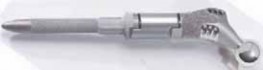
During the past decade remarkable advances in the field of revision hip reconstruction have been made. One such improvement has been the introduction of second-generation modular prosthetic components ( FIG 1), which provide improved ability to restore limb length and achieve optimal soft tissue tension, both of which may reduce the incidence of instability that often followed insertion of a monolithic megaprosthesis. A new generation of megaprostheses also provides a better environment for soft tissue reattachment and the ability to reapproximate the retained host bone to the prosthesis. ANATOMY
3.
The abductors are the gluteus medius and minimus muscles, the tensor fascia lata muscles, and the iliotibial band. The adductors are the adductor brevis, adductor longus, and gra-cilis muscles, and the anterior part of the adductor magnus muscle. The external rotators are the piriformis, quadratus femoris, superior gemellus, inferior gemellus, obturator inter-nus, and obturator externus muscles.
4.
Abductors are important stabilizers of the hip that are inner-vated mainly by the superior gluteal nerve. The nerve exits the pelvis via the suprapiriform portion of the sciatic foramen along with the superior gluteal vessels. Palsy results in abductor lurch, a Trendelenburg gait. PATHOGENESIS
5.
Femoral bone loss is a constantly rising, predominantly complex and challenging problem in revision arthroplasty.
6.
Numerous factors may contribute to the loss of femoral bone stock encountered in revision hip arthroplasty:
1.
Osteolysis secondary to particle debris
2.
Stress-shielding with adaptive bone-remodeling
3.
Previous infection
 ---
---
FIG 1 • New generation of modular proximal femoral and total femoral replacement prostheses (Stryker Orthopedics, Mahwah, NJ).
-
Natural processes of aging
8.
Periprosthetic fracture
1.
Multiple previous failed reconstructive procedures with insertion and removal of implants also compromise the integrity of the bone stock and adversely affect the integrity and function of the abductor muscles.
2.
Options available for dealing with severe femoral bone loss include the use of a long cemented stem or press-fit stems, im-paction allografting, resection arthroplasty, allograft prosthetic replacement, and modular replacement.
3.
An allograft–prosthesis composite potentially increases bone stock in the proximal part of the femur and provides sites for soft tissue attachment, including the abductor muscles. The use of an allograft–prosthesis composite is limited due to the risks of infection, junctional nonunion, dislocation, and aseptic loosening.
4.
A proximal femoral replacement probably is more available to most surgeons than a proximal femoral allograft, and it is less technically demanding to implant.
5.
Zehr et al28 conducted a comparative study in which the use of an allograft–prosthesis composite was found to be marginally better than proximal femoral replacement for reconstruction after tumor resection. NATURAL HISTORY
6.
The megaprosthesis is valuable in the armamentarium of the reconstructive hip surgeon who treats patients with extensive bone loss for whom other available reconstructive procedures cannot be utilized.
7.
This prosthesis will have an unacceptably high failure rate in younger patients, and other reconstructive options should be explored. PATIENT HISTORY AND PHYSICAL FINDINGS
8.
Assessment of the present and past medical history, physical examination results, and radiographic findings lead to a correct diagnosis of hip pathology in most patients.
9.
Intra-articular or acetabular pathology usually presents as groin pain.
10.
Thigh pain (especially start-up pain) is more indicative of a loose femoral stem.
11.
Patients may present with severe knee pain as a result of hip pathology. Any patient with knee pain should be evaluated for hip pathology.
12.
Neurovascular examination and examination of the spine and abdomen should be conducted to exclude other reasons of hip pain, such as neuropathies, vascular claudication, and spinal stenosis.
1.
Specific tests for the hip with evaluation of range of motion and leg lengths should be documented.
1.
Leg-length assessment for apparent or functional leg length discrepancy: a deficiency may be due to pelvic obliq-uity, contractures, or scoliosis.
2.
Trendelenburg test: inability to stabilize the pelvis indicates abductor weakness.
3.
Straight leg raise: radicular pain may be felt along the leg.
4.
Thomas test: evaluates for hip flexion contracture.
5.
Stinchfield test: groin pain or weakness may indicate intra-articular hip pathology.
2.
Initial history taking should begin with a discussion of the patient’s chief complaint. The location and nature of the pa-tient’s pain can guide the surgeon to the proper diagnosis.
3.
A thorough review of the patient’s medical history along with a complete review of systems will help the surgeon to identify any potential factors that may lead to perioperative complications and provides an opportunity to medically treat or optimize the patient before the planned operation.
4.
Sources of potential or concurrent infection must be discovered, and proper evaluation and treatment should be performed well in advance of the surgical procedure.
5.
Negative hip aspirations do not completely rule out infection and should be followed up with intraoperative tissue sampling with frozen sections after alerting the appropriate pathology department personnel well before the planned surgical date.
6.
Patients with any history of chronic venous stasis ulcers, previous vascular bypass surgery, or absent distal pulses should be evaluated by a vascular surgeon.
7.
The physical examination should begin with the analysis of the patient’s gait. Use of ambulatory assistive devices, a limp, or a deformity of the lower extremity should be noted.
8.
The antalgic gait is a result of pain in all phases of ambulation with weight bearing and is characterized by a shortened stance phase indicating hip-joint disease.
9.
The Trendelenburg gait or abductor lurch indicates either paralysis or loss of continuity of the abductor musculature and is identified by observing the shift of the patient’s center of gravity over the affected extremity during the stance phase of gait.
10.
Inspection of previous surgical wounds should be routinely performed. Planning of the surgical incision is important in determining the approach for the surgical reconstruction, and, although skin flap necrosis after hip surgery is rare, the maximum distance and angles used should be optimal to avoid this complication.
11.
The active and passive ranges of motion of the hip should be identified along with the strength of the hip girdle musculature. IMAGING AND OTHER DIAGNOSTIC STUDIES
12.
Proximal and total femur resections are major surgical procedures that necessitate a detailed preoperative evaluation.
13.
Physical examination and imaging studies aid in determination of the extent of bone resection and dimensions of the required prosthesis; the extent of soft tissue resection and reconstruction possibilities; and the proximity of the scarred-in femoral vessels, femoral nerve, and sciatic nerve.
14.
Most complications can be avoided by anticipating them before surgery and modifying the surgical technique accordingly.
15.
Plain radiographs are used to evaluate the extent and level of bone destruction. If needed, CT scanning can be added for further delineation of the femur and acetabulum bone structure.
16.
MRI is used to evaluate the medullary canal and soft tissue around the hip joint.
17.
Three-phase bone scan is essential to determine the presence of metastatic bone disease.
18.
Angiography of the iliofemoral vessels is essential before proximal femoral replacement if distortion of the anatomy following multiple previous surgeries is suspected. DIFFERENTIAL DIAGNOSIS
19.
Osteomyelitis
20.
Metastatic lesions
21.
Primary bone tumors, eg, multiple myeloma, chondro-sarcoma
22.
Periprosthetic fracture
23.
Osteolysis
24.
Aseptic loosening
25.
Paget disease
26.
Metabolic disease NONOPERATIVE MANAGEMENT
27.
For the indications discussed in this chapter, the only reasonable option would be a surgical one. If the patient’s medical problems are serious enough to put off the surgery, however, an antidislocation brace with protected weight bearing can be used. SURGICAL MANAGEMENT
28.
Use of the megaprosthesis (ie, proximal femoral replacement and total femur replacement) is reserved to expedite recovery for elderly or sedentary patients with massive bone loss that may have occurred after failed total hip arthroplasty ( FIG 2A,B), deep infection, periprosthetic fracture (FIG 2C,D), fracture nonunion with failed multiple attempts at osteosynthe-sis, and hip salvage after a failed resection arthroplasty.
29.
In younger patients with bone loss of high magnitude that cannot be reconstructed by conventional means, an allograft prosthetic composite is preferred over femoral prosthetic replacement.
30.
An important prerequisite for the use of prosthetic femoral replacement and allograft prosthetic composite is the availability of sufficient distal femoral length (less than 10 cm) for secure fixation of the cemented or uncemented femoral stem.
31.
When distal bone is severely deficient, total femoral replacement may be considered.
32.
The presence of superficial or deep infection around the hip is an absolute contraindication.
33.
Additional contraindications include lack of cooperation on the part of the patient, vascular insufficiency that may prevent healing, and the presence of significant medical comorbidities precluding administration of anesthesia. Preoperative Planning
34.
The importance of preoperative planning in hip arthroplasty in general, and in proximal femur reconstruction in particular, cannot be overstated. These cases can be technically demanding, requiring meticulous attention to detail to achieve success.
 ---
---
 ---
---

 ---
---
 ---
A B C D ### FIG 2 • A. AP radiograph of a patient with multiple previous surgeries for deep infection that had resulted in massive proximal femoral bone loss. B. A megaprosthesis was used for reconstruction. C. A 72-year-old patient presenting with periprosthetic fracture. D. Because of severe bone loss, reconstruction with megaprosthesis was carried out.
1.
Proximal femur reconstruction is performed for metaphy-seal–diaphyseal lesions that extend below the lesser trochanter, cause extensive cortical destruction, and spare at least 3 cm of the distal femoral diaphysis.
2.
Total femur resection is performed for diaphyseal lesions that extend proximally to the lesser trochanter and distally to the distal diaphyseal–metaphyseal junction and cause extensive bone destruction.
3.
Preoperative clinical and radiographic (standing films) assessment of limb length are carried out and recorded.
4.
Intraoperative monitoring of the sciatic and femoral nerves may be required in patients in whom extensive limb lengthening (more than 4 cm) is anticipated.
5.
Preoperative templating to select the appropriate stem length and diameter is essential.
6.
Problems with removal of existing hardware, specific needs for acetabular reconstruction, the potential need for insertion of constrained liners, and determining the absence of previous infection should be anticipated and addressed appropriately.
7.
Even with the most accurate preoperative measurements, a variety of prosthesis sizes should be available in the operating room, because intraoperative adjustments with change in the anticipated size of the prosthesis are common.
8.
The representative of the company that manufactured the prosthesis to be used in the proximal femur reconstruction should be present in the operating room.
9.
The operating room personnel, particularly the scrub person, who assist with this procedure should be experienced. An experienced anesthesia team should administer anesthesia, because these patients often are elderly and frail, and invasive monitoring often is warranted.
10.
Regional anesthesia is preferred. Intraoperative blood salvage (ie, with a cell saver) should be used in these patients.
11.
The anesthesia team should be prepared for the possibility of large volume loss and encouraged to monitor this closely.
12.
Invasive monitoring with arterial lines or pulmonary catheters may be necessary in some patients. Positioning
1.
We place the patient in the lateral decubitus or supine position.
2.
Nonpermeable U-drapes are used to isolate the groin.
3.
The distal third of the extremity also is isolated from the field using impermeable drapes. The knee must be included in the operative field, even in patients undergoing proximal femoral replacement.
4.
Extension of the incision and arthrotomy of the knee to address intraoperative problems such as fractures extending distally is not uncommon.
5.
The skin is scrubbed with povidone-iodine solution for at least 10 minutes and DuraPrep (3M, St. Paul, MN) applied before application of Ioban (3M) to the skin. Approach
6.
We use the direct lateral approach (Hardinge approach) or the posterolateral approach with trochanteric slide osteotomy to gain access to the hip and maintain a low threshold to extend the incision as needed ( FIG 3).
---
A B C D ### FIG 2 • A. AP radiograph of a patient with multiple previous surgeries for deep infection that had resulted in massive proximal femoral bone loss. B. A megaprosthesis was used for reconstruction. C. A 72-year-old patient presenting with periprosthetic fracture. D. Because of severe bone loss, reconstruction with megaprosthesis was carried out.
1.
Proximal femur reconstruction is performed for metaphy-seal–diaphyseal lesions that extend below the lesser trochanter, cause extensive cortical destruction, and spare at least 3 cm of the distal femoral diaphysis.
2.
Total femur resection is performed for diaphyseal lesions that extend proximally to the lesser trochanter and distally to the distal diaphyseal–metaphyseal junction and cause extensive bone destruction.
3.
Preoperative clinical and radiographic (standing films) assessment of limb length are carried out and recorded.
4.
Intraoperative monitoring of the sciatic and femoral nerves may be required in patients in whom extensive limb lengthening (more than 4 cm) is anticipated.
5.
Preoperative templating to select the appropriate stem length and diameter is essential.
6.
Problems with removal of existing hardware, specific needs for acetabular reconstruction, the potential need for insertion of constrained liners, and determining the absence of previous infection should be anticipated and addressed appropriately.
7.
Even with the most accurate preoperative measurements, a variety of prosthesis sizes should be available in the operating room, because intraoperative adjustments with change in the anticipated size of the prosthesis are common.
8.
The representative of the company that manufactured the prosthesis to be used in the proximal femur reconstruction should be present in the operating room.
9.
The operating room personnel, particularly the scrub person, who assist with this procedure should be experienced. An experienced anesthesia team should administer anesthesia, because these patients often are elderly and frail, and invasive monitoring often is warranted.
10.
Regional anesthesia is preferred. Intraoperative blood salvage (ie, with a cell saver) should be used in these patients.
11.
The anesthesia team should be prepared for the possibility of large volume loss and encouraged to monitor this closely.
12.
Invasive monitoring with arterial lines or pulmonary catheters may be necessary in some patients. Positioning
1.
We place the patient in the lateral decubitus or supine position.
2.
Nonpermeable U-drapes are used to isolate the groin.
3.
The distal third of the extremity also is isolated from the field using impermeable drapes. The knee must be included in the operative field, even in patients undergoing proximal femoral replacement.
4.
Extension of the incision and arthrotomy of the knee to address intraoperative problems such as fractures extending distally is not uncommon.
5.
The skin is scrubbed with povidone-iodine solution for at least 10 minutes and DuraPrep (3M, St. Paul, MN) applied before application of Ioban (3M) to the skin. Approach
6.
We use the direct lateral approach (Hardinge approach) or the posterolateral approach with trochanteric slide osteotomy to gain access to the hip and maintain a low threshold to extend the incision as needed ( FIG 3).
 ---
---
FIG 3 • Diagram showing the placement of incision.
TECHNIQUES EXPOSURE 1. When extensile exposure of the femur is needed, a vastus slide osteotomy, as described by Head et al,12 mobilizes the anterior abductor, vastus lateralis, and vastus inter-medius muscles anteriorly in unison and exposes the anterior and lateral aspects of the femur ( TECH FIG 1). 2. Meticulous soft tissue handling helps the tissues to heal and minimizes postoperative complications. 3. Deep tissue specimens for frozen section and culture are obtained in all cases. Meticulous débridement of the hip is carried out to remove previous metal debris and hardware around the femur, if present. 4. When a posterolateral incision is routinely used for proximal femur resections, the incision can be extended to the anterolateral aspect of the patellar tendon if a total femur resection is required. 5. The abductors are identified, as are their anterior and posterior intervals. The abductors are transected through their tendinous attachments and retracted, exposing the hip joint and acetabulum. 6. The vastus lateralis is reflected distally from its origin, and the posterior perforating vessels are ligated. The vastus lateralis has to be preserved because of its future role in soft tissue coverage of the prosthesis; it will be advanced proximally and sutured to the abductors. 1. Care is taken not to ligate its main pedicle, which crosses anteriorly and obliquely along the rectus femoris fascia.
 --- TECH FIG 1 • Exposure of the femur in a patient who has sustained a periprosthetic fracture. PROXIMAL FEMORAL REPLACEMENT 1. If the femur is intact, an osteotomy to split the proximal femur may be required to facilitate removal of the previous prosthesis or hardware. 2. A transverse osteotomy first is made in the host bone at the most proximal area of bone with good circumferen-tial quality. 3. Because the outcome of this procedure is influenced directly by the length of the remaining femur, maximum length of the native femur is maintained at all costs.16 4. We then use a longitudinal Wagner type of coronal plane osteotomy to split the proximal femur if the bone quality is poor. ❚ 5. Soft tissue attachments to the proximal femur— particularly the abductor mechanism, if present—should be retained if at all possible. Once the femur is exposed, the distal portion of the canal is prepared by successive broaching. The cancellous bone, when present, is preserved for better cement interdigitation. 6. After completion of femoral preparation and determination of the size of best-fit broach, trial components are inserted, and the stability of the hip is examined. 7. A distal cement restrictor is used whenever possible. The restrictor is introduced and advanced distally to allow for at least 2 cm of bone cement at the tip of the stem. 8. The cement is pressurized and the final component implanted, with care taken to ensure that the porous coated portion of the stem is placed directly and firmly against diaphyseal bone with no interpositioning cement. 9. The prosthesis can be assembled and then cemented distally or, alternatively, the stem can be cemented and then the body assembled onto it. 1. Extreme care must be exercised to prevent rotational malpositioning ( TECH FIG 2). To mark the rotation, we use a sharp osteotome to scratch the distal femoral cortex once the trial component is appropriately positioned. The rotation of the component cannot be changed once the distal stem is cemented in place.
 --- TECH FIG 2 • Demonstration of how the rotational positioning or version of the femoral component is determined. The version of the femoral stem is judged by appropriate positioning of the knee. #### TOTAL FEMUR REPLACEMENT
- Indications for total femoral replacement are rare and generally include inadequate length (less than 10 cm) or such poor quality of distal femoral bone that fixation of a femoral stem is precluded. In most cases, the distal femur is of adequate length and quality to allow secure fixation. 2. Total femoral replacement includes an arthrotomy of the knee to allow prosthetic replacement of the knee. 3. Once exposure of the femur is completed using a lateral vastus reflecting approach, the entire femur is split lon-gitudinally in the coronal plane. 4. Again, even if it is of extremely poor quality, as much of the bone with its soft tissue attachment as possible is retained. 5. The subvastus approach is extended to include a lateral or medial arthrotomy of the knee and eversion of the patella. 6. The amount of tibial bone resected is kept to a minimum, but it must be of adequate thickness to allow implantation of the components and insertion of polyethylene without elevating the joint line. The tibia is prepared in the same manner as for total knee arthroplasty. Once appropriate tibial component size is determined, preparation of the tibia followed by insertion of the trial component is carried out. TECHNIQUES 1. A full-length trial femur is assembled, ensuring that appropriate limb length is restored. Unless constrained liners are to be used, we prefer to use a large femoral head size to improve arc of motion and minimize instability. 2. The tibial polyethylene usually is between 15 and 20 mm thick, but it may be necessary to adjust the thickness to obtain appropriate length of the extremity and restore the joint line. 3. A linked articulated knee design is necessary because of loss of the stabilizing ligamentous structures. Once the prosthesis is assembled, a trial reduction is carried out and tested for stability. 4. We usually do not resurface the patella unless severe wear of the articular cartilage is noted. #### DETERMINATION OF LIMB LENGTH
- The length of the femoral component is determined through careful preoperative planning and intraoperative assessment. 2. Two methods may be used for proper leg length determination. 1. The first method is to apply traction to the limb with measurement from the cup to the host bone osteotomy site (for proximal femoral replacement). 2. The second and preferred method is to place a Steinmann pin in the iliac crest to measure a fixed point on the femur before dislocation. 3. With the long-stem trial prosthesis in place, proper leg length can be accurately restored. For patients with total femur replacement, radiographs of the opposite, normal femur may be obtained preoperatively and used for accurate templating for length. 5. The length of the prosthesis usually equals the length of the bone being resected, although in many patients the integrity of the bone has been breached and the anatomy markedly altered. 6. Ultimately, the femoral prosthesis length depends on the soft tissue tension about the hip. Balancing tension, restoration of limb length, and avoiding excessive tension on the sciatic nerve are of utmost importance if complications are to be avoided. #### ACETABULAR RECONSTRUCTION
-
The acetabulum is exposed at the beginning of the operation and examined carefully. If a previous acetabular component is in place, the stability and positioning of the component are scrutinized.
2.
If the component is appropriately placed and stable, it is left in place, and the liner is exchanged. If a previous acetabular component is not in place, a new component is inserted in a press-fit manner with screw fixation.
3.
More complex acetabular reconstruction, eg, the use of an antiprotrusio cage, occasionally is needed.
4.
The type of acetabular liner is determined after reconstruction of the femur has been completed, because it may be necessary to use constrained liners in patients with poor soft tissue tension and a high probability of instability.
7.
The constrained liners can be either snap-fit or cemented into the shell, depending on the type of the acetabular component implanted. In our experience, constrained liners are required in approximately half of patients receiving a megaprosthesis.
8.
Our absolute indication for the use of a constrained liner is for patients with properly positioned components and equal or near equal leg length who have intraoperative instability secondary to soft tissue deficiency.
 ---
---


 ---
---
 ---
---
