العربية

Revision THA: Acetabular Impaction Grafting for Severe Bone Loss

Updated: Feb 2026 43 Views
Revision THA: Acetabular Impaction Grafting for Severe Bone Loss
📖 Clinical Article

Case Title: Acetabular Impaction Grafting

Demographics
1. Age: 79 Sex: female BMI: 22
2. #### Relevant Past Medical History
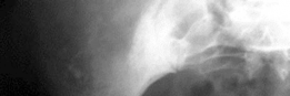
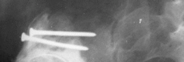
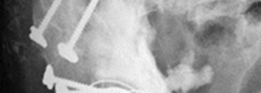
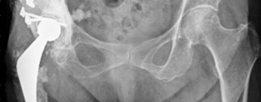
Principal pathologies : hip ankylosis in 1954 due to DDH (Fig. 18.5).
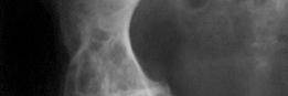
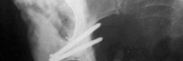
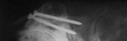
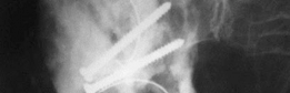
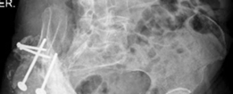
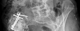
Previous surgical procedures : primary and acetabular revision right THA in 1974 (Fig. 18.6). History of Presenting Complaint : right hip pain.
3. #### Clinical Examination
Symptoms: pain
Range of motion: severely altered
Main disability: unable to walk more than one hundred metres
Scoring if available: Merle DÁubigne 6 out of 18 points
Neurovascular evaluation: normal
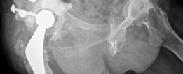
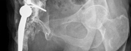
4. Preoperative Radiological Assessment/Imaging(Figs. 18.5, 18.6, and 18.7)

Illustration 1 for Revision THA: Acetabular Impaction Grafting for Severe Bone Loss
🔍 Click to enlarge
Clinical Radiograph / Orthopedic Image

Illustration 2 for Revision THA: Acetabular Impaction Grafting for Severe Bone Loss
🔍 Click to enlarge
Clinical Radiograph / Orthopedic Image

Illustration 3 for Revision THA: Acetabular Impaction Grafting for Severe Bone Loss
🔍 Click to enlarge
Clinical Radiograph / Orthopedic Image

Illustration 4 for Revision THA: Acetabular Impaction Grafting for Severe Bone Loss
🔍 Click to enlarge
Clinical Radiograph / Orthopedic Image
Fig. 18.5 Anteroposterior right hip radiograph of the patient in 1974, when a right Charnley total hip arthroplasty was indicated
Illustration 5 for Revision THA: Acetabular Impaction Grafting for Severe Bone Loss
🔍 Click to enlarge
Clinical Radiograph / Orthopedic Image

Illustration 6 for Revision THA: Acetabular Impaction Grafting for Severe Bone Loss
🔍 Click to enlarge
Clinical Radiograph / Orthopedic Image

Illustration 7 for Revision THA: Acetabular Impaction Grafting for Severe Bone Loss
🔍 Click to enlarge
Clinical Radiograph / Orthopedic Image

Illustration 8 for Revision THA: Acetabular Impaction Grafting for Severe Bone Loss
🔍 Click to enlarge
Clinical Radiograph / Orthopedic Image
Fig. 18.6 Anteroposterior right hip radiograph showing a Charnley total hip arthroplasty with two cemented cups, the upper one was first cemented, forty eight hours later the patient was sent to the operating room again, and a new cemented cup in the proper position was implanted
Illustration 9 for Revision THA: Acetabular Impaction Grafting for Severe Bone Loss
🔍 Click to enlarge
Clinical Radiograph / Orthopedic Image

Illustration 10 for Revision THA: Acetabular Impaction Grafting for Severe Bone Loss
🔍 Click to enlarge
Clinical Radiograph / Orthopedic Image

Illustration 11 for Revision THA: Acetabular Impaction Grafting for Severe Bone Loss
🔍 Click to enlarge
Clinical Radiograph / Orthopedic Image

Illustration 12 for Revision THA: Acetabular Impaction Grafting for Severe Bone Loss
🔍 Click to enlarge
Clinical Radiograph / Orthopedic Image
Fig. 18.7 Anteroposterior right hip radiograph depicting acetabular loosening and massive bone loss thirty four years postoperatively. We indicated revision hip surgery with a trabecular metal cup and augment 5. #### Preoperative Planning
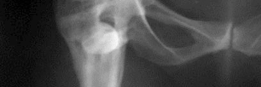
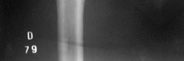
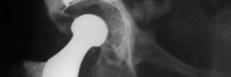
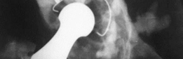
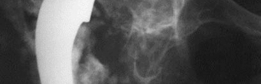
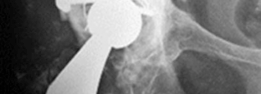
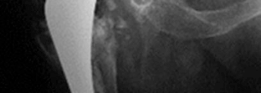
Diagnosis : acetabular loosening of a right THA (Fig. 18.7).
Possible treatment options: revision THA.
Chosen treatment method: bone reconstruction with strut and cancellous impaction grafting.
Selection of implants if applicable and rational : we planned to use a jumbo tantalum cup, but due to the amount of bone loss, that cup was not possible to be implanted (Figs. 18.8 and 18.9).
Strategies to overcome difficulties : we tailored an allograft belonging to a proximal femur and impacted it between the anterior and posterior columns; then we fixed it with two screws to the iliac bone and two screws to the ischium, and then we cemented a cup (Figs. 18.9, 18.10, 18.11,
18.12, 18.13 and 18.14).
Illustration 13 for Revision THA: Acetabular Impaction Grafting for Severe Bone Loss
🔍 Click to enlarge
Clinical Radiograph / Orthopedic Image

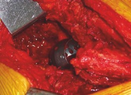
--- Fig. 18.8 Intraoperative view showing the massive bone loss as determined by the number 52 reamer
Illustration 14 for Revision THA: Acetabular Impaction Grafting for Severe Bone Loss
🔍 Click to enlarge
Clinical Radiograph / Orthopedic Image

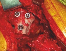
--- Fig. 18.9 A proximal femur was tailored to be impacted between the anterior and posterior column of the acetabulum and then fixed with four screws, two directed to the iliac bone and two to the ischium. After achieving stability of the strut, the morselized bone graft was impacted in the medial wall. The strut allograft was reamed, and some holes were performed for cement penetration
Illustration 15 for Revision THA: Acetabular Impaction Grafting for Severe Bone Loss
🔍 Click to enlarge
Clinical Radiograph / Orthopedic Image

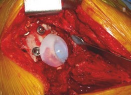
--- Fig. 18.10 An Ogee cemented cup was cemented, and femoral reconstruction was performed with impaction grafting and a long cemented stem 6. #### Surgical Note
Patient’s position: lateral decubitus.
Type of anaesthesia: hypotensive epidural.
Surgical approach: posterolateral.
Main steps: see above.
Reconstruction techniques: see above.
7. #### Intraoperative Challenges
Challenges and solutions : massive bone defect, unable to be reconstructed with trabecular metal technology (Fig. 18.8).
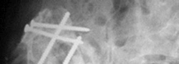
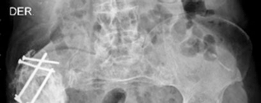
8. #### Postoperative Radiographs
(Figs. 18.11, 18.12, 18.13,
and 18.14)
Illustration 16 for Revision THA: Acetabular Impaction Grafting for Severe Bone Loss
🔍 Click to enlarge
Clinical Radiograph / Orthopedic Image

Illustration 17 for Revision THA: Acetabular Impaction Grafting for Severe Bone Loss
🔍 Click to enlarge
Clinical Radiograph / Orthopedic Image

Illustration 18 for Revision THA: Acetabular Impaction Grafting for Severe Bone Loss
🔍 Click to enlarge
Clinical Radiograph / Orthopedic Image

Illustration 19 for Revision THA: Acetabular Impaction Grafting for Severe Bone Loss
🔍 Click to enlarge
Clinical Radiograph / Orthopedic Image
Fig. 18.11 Immediate postoperative radiographs showing the anatomical centre of rotation
Illustration 20 for Revision THA: Acetabular Impaction Grafting for Severe Bone Loss
🔍 Click to enlarge
Clinical Radiograph / Orthopedic Image

Illustration 21 for Revision THA: Acetabular Impaction Grafting for Severe Bone Loss
🔍 Click to enlarge
Clinical Radiograph / Orthopedic Image
Fig. 18.12 Anteroposterior both hips radiograph at seven years’ follow-up
Illustration 22 for Revision THA: Acetabular Impaction Grafting for Severe Bone Loss
🔍 Click to enlarge
Clinical Radiograph / Orthopedic Image

Illustration 23 for Revision THA: Acetabular Impaction Grafting for Severe Bone Loss
🔍 Click to enlarge
Clinical Radiograph / Orthopedic Image
Fig. 18.13 Oblique obturator view at seven years’ follow-up
Illustration 24 for Revision THA: Acetabular Impaction Grafting for Severe Bone Loss
🔍 Click to enlarge
Clinical Radiograph / Orthopedic Image

Illustration 25 for Revision THA: Acetabular Impaction Grafting for Severe Bone Loss
🔍 Click to enlarge
Clinical Radiograph / Orthopedic Image
Fig. 18.14 Oblique iliac view at seven years’ follow-up 9. #### Postoperative Management
Chemoprophylaxis and anticoagulant treatment period: enoxaparin for four weeks.
Gait/limb loading until full loading: three months of non-weight bearing and three months of partial weight bearing, then full weight bearing.
10. #### Follow-Up and Complications
Scoring: 14 points Merle DÁubigne score.
11. Discussion[26, 39, 40]
Advantages of the applied method: bone reconstruction.
Disadvantages of the method: old patient.
Alternative evidence-based techniques for the case: no alternatives.
Why is the chosen technique better for this case: the only one we found.
Indications and contraindications for your technique: indication is the bone loss and contraindication is the active infection.
Learning curve and how to manage complications: prolonged learning curve.
Level of evidence concerning the superiority of this method against others: low level.

Dr. Mohammed Hutaif
Written & Medically Reviewed by
Consultant Orthopedic & Spine Surgeon