Introduction & Epidemiology
The forearm, a functionally critical segment of the upper extremity, is comprised of the radius and ulna, articulated proximally by the proximal radioulnar joint (PRUJ) and distally by the distal radioulnar joint (DRUJ). Its unique anatomy, characterized by the inherent curvature of the radial diaphysis – the radial bow – is fundamental for the complex kinematics of pronation and supination. This elegant anatomical feature allows the radius to rotate around a relatively fixed ulnar axis, creating a stable yet highly mobile platform for hand function. Disruption of this radial bow, whether through acute trauma leading to fracture, malunion, or iatrogenic miscontouring during osteosynthesis, inevitably compromises forearm rotation and can severely impair overall upper limb function.
Forearm fractures, particularly diaphyseal fractures involving both the radius and ulna, are common injuries, accounting for approximately 10-15% of all adult fractures. They represent a significant challenge due to the high biomechanical demands placed on the forearm and the critical need to restore precise anatomical relationships. In adults, these fractures typically result from high-energy trauma, often leading to significant displacement and instability. The incidence exhibits a bimodal distribution, affecting young, active individuals and the elderly, with distinct injury mechanisms and associated comorbidities. In younger cohorts, sports injuries and motor vehicle accidents are prevalent etiologies, while falls are more common in older populations.
The primary objective in managing diaphyseal forearm fractures is the restoration of forearm length, axial alignment, and most critically, the intrinsic radial bow. Failure to achieve anatomical reduction, particularly the restoration of the radial length and curvature, leads to malunion, which can manifest as persistent pain, restriction of pronation/supination, and ultimately, functional disability. This comprehensive review aims to delineate the intricate surgical anatomy and biomechanics of the radial bow, outline contemporary operative strategies, and discuss potential complications and their management, underscoring the paramount importance of anatomical restoration for optimal functional outcomes.
Surgical Anatomy & Biomechanics
The radial bow is a characteristic curvature of the radial shaft, typically exhibiting both a dorsal and a lateral concavity. This double-curved morphology is essential for maintaining the interosseous space and facilitating the intricate rotational mechanics of the forearm.
The radius is convex dorsally in its proximal third, relatively straight in its middle third, and then convex ventrally in its distal third. Laterally, the radius has a gentle curve, being convex laterally in its mid-diaphysis. These curvatures ensure that the radius maintains sufficient distance from the ulna throughout the arc of pronation and supination, preventing impingement of the interosseous membrane and the surrounding musculature.
Key Anatomical Features:
*
Radial Diaphysis:
The shaft of the radius exhibits a distinct posterior and lateral bow. The average dorsal bow is approximately 10-15 degrees, with the apex typically located around the junction of the proximal and middle thirds. The lateral bow is less pronounced but equally critical.
*
Ulna:
In contrast, the ulna is relatively straight, acting as the stable axis around which the radius rotates. The olecranon and coronoid processes articulate with the humerus, forming the hinge-like humeroulnar joint, which contributes to elbow stability.
*
Interosseous Membrane (IOM):
A fibrous sheet connecting the radius and ulna, the IOM plays a vital role in longitudinal stability, load sharing between the two bones, and facilitating muscle attachment. Its fibers are oriented obliquely, largely responsible for transmitting axial forces from the radius to the ulna. Preservation or reconstruction of the IOM's integrity is critical for preventing proximal migration of the radius and maintaining forearm stability.
*
Proximal Radioulnar Joint (PRUJ):
The radial head articulates with the radial notch of the ulna, secured by the annular ligament. This trochoid joint allows for the pivotal rotation of the radial head.
*
Distal Radioulnar Joint (DRUJ):
The distal radius articulates with the ulnar head, stabilized by the triangular fibrocartilage complex (TFCC). The TFCC is crucial for DRUJ stability, load transmission across the wrist, and smooth pronation-supination.
Biomechanics of Forearm Rotation:
Pronation and supination are complex motions that involve coordinated movement at both the PRUJ and DRUJ. During these movements, the radial head rotates within the annular ligament at the PRUJ, while the distal radius circumducts around the relatively fixed ulnar head at the DRUJ.
The radial bow is fundamental to this process for several reasons:
1.
Maintenance of Interosseous Space:
The curvature creates a crucial space between the radius and ulna, accommodating the bulk of the forearm muscles and preventing soft tissue impingement during rotation. Without this bow, the muscle mass would be compressed, impeding motion.
2.
Optimal DRUJ Kinematics:
The precise curvature dictates the path of the distal radius around the ulna. Any alteration in the radial bow (e.g., increased straightness, excessive angulation) significantly alters the DRUJ mechanics, leading to impingement, restricted motion, and potentially early degenerative changes.
3.
Mechanical Advantage:
The bow provides optimal lever arms for the pronator and supinator muscles, maximizing their efficiency.
4.
Load Sharing:
While the IOM is the primary load-sharing structure, the overall architectural integrity, including the radial bow, contributes to the appropriate distribution of axial forces across the forearm, wrist, and elbow.
Restoration of the radial bow typically involves addressing three-dimensional parameters: length, rotation, and angulation. Each degree of rotational or angular malalignment can significantly compromise forearm function. A loss of the radial bow, often seen in malunited diaphyseal fractures, results in decreased interosseous space, leading to abutment between the radius and ulna during rotation. This can manifest as a restriction in pronation and supination, often with a compensatory increase in shoulder and wrist motion, and can cause chronic pain due to impaction or impingement. Therefore, precise anatomical reduction and rigid internal fixation are paramount to restore the native radial bow and preserve unimpeded forearm rotation.
Indications & Contraindications
Management of forearm fractures, especially those involving the radial diaphysis, is overwhelmingly surgical in adults due to the high rates of malunion, nonunion, and functional impairment associated with non-operative treatment of displaced fractures.
Indications for Operative Management:
-
Adult Diaphyseal Forearm Fractures:
- Displaced Radial Shaft Fractures: Any significant angulation (>10 degrees), translation (>50%), or shortening (>5mm) of the radial shaft.
- Both-Bone Forearm Fractures (Radius and Ulna): The vast majority of these in adults require open reduction and internal fixation (ORIF) of both bones to achieve stable fixation and restore anatomy.
- Galeazzi Fracture-Dislocation: Fracture of the radial shaft with associated DRUJ dislocation/subluxation. Always operative in adults to stabilize the radius and indirectly reduce the DRUJ.
- Monteggia Fracture-Dislocation (Adult): Fracture of the ulnar shaft with associated radial head dislocation. Operative fixation of the ulna is critical to reduce the radial head.
- Open Fractures: Require urgent debridement and stabilization.
- Neurovascular Compromise: Urgent operative exploration and stabilization.
- Impending Compartment Syndrome: Prophylactic fasciotomy and stabilization.
- Malunion of Forearm Fractures: Symptomatic malunion leading to significant loss of pronation/supination, pain, or cosmetic deformity. Corrective osteotomy is indicated.
- Nonunion of Forearm Fractures: Diagnosed after 6-9 months without radiographic evidence of healing, especially if symptomatic or unstable. Requires revision ORIF, often with bone grafting.
- Pathological Fractures: Due to tumors or metabolic bone disease.
- Floating Elbow Injuries: Coexistent elbow and forearm fractures.
Indications for Non-Operative Management:
- Minimally Displaced or Stable Undisplaced Fractures: Extremely rare for adult diaphyseal forearm fractures to be treated non-operatively, given the high risk of malunion. This might be considered only in very select, stable, non-displaced greenstick or buckle fractures, often in skeletally immature patients.
- Pediatric Forearm Fractures: Many pediatric forearm fractures can be managed non-operatively with closed reduction and casting due to their greater remodeling potential. However, significant displacement, instability, or loss of radial bow in older children may still warrant operative intervention.
- Medically Unfit Patients: Patients with severe comorbidities precluding safe surgical intervention, where the risks of surgery outweigh the potential benefits. This often necessitates accepting a compromised functional outcome.
- Palliative Care Settings: For patients with very limited life expectancy or severe underlying neurological conditions where functional recovery is not a primary goal.
Summary of Indications
| Indication Type | Operative Management Summary of Key Literature / Guidelines
The principles governing the restoration of the radial bow are deeply rooted in understanding forearm biomechanics, first emphasized by the seminal work of Böhler and subsequently elaborated upon by the AO Foundation. The overarching directive remains that surgical intervention for displaced adult forearm fractures, particularly those involving the radius, is mandatory to prevent functional loss due to malunion or nonunion.
- AO Principles: The Arbeitsgemeinschaft für Osteosynthesefragen (AO) has long championed the meticulous anatomical reduction and stable internal fixation of both bones in adult forearm fractures. Their guidelines emphasize the importance of restoring length, rotation, and axial alignment, which inherently means restoring the radial bow. Plate osteosynthesis with dynamically compressing plates (DCP) or limited-contact dynamic compressing plates (LC-DCP) has been the gold standard, providing rigid fixation allowing for early mobilization. More recently, locking compression plates (LCP) have gained prominence, offering angular stability particularly beneficial in osteoporotic bone or comminuted fractures where absolute stability might be challenging with conventional plates.
- Plate Choice and Contouring: While pre-contoured plates are increasingly available, they may not perfectly match every patient's unique radial bow. Surgeons must understand the importance of meticulous plate contouring to replicate the natural curvature of the radius. Over-contouring or under-contouring can both lead to functional deficits. Studies by Scheer et al. have quantitatively described the radial bow, providing anatomical data to guide surgical reconstruction.
- Minimum Fixation Requirements: Current literature supports the use of at least six cortices (three screws) proximally and distally to the fracture site for robust fixation. For comminuted fractures or in osteoporotic bone, longer plates and more screws may be necessary. Bicortical screw fixation is generally preferred for diaphyseal forearm fractures to achieve maximum stability.
- Both-Bone Fixation: While fixation of one bone can sometimes reduce the other, especially in children, current adult practice dictates plating of both radius and ulna when both are fractured and displaced. This ensures precise restoration of the interosseous relationship and prevents synostosis.
- Timing of Surgery: Early surgical intervention for acute displaced forearm fractures is generally associated with better outcomes, reducing soft tissue swelling and facilitating easier reduction. Delay in surgery can increase the risk of complications such as compartment syndrome, infection, and chronic pain.
- Management of Malunion: Symptomatic malunion, particularly with loss of radial bow, is a challenging problem often requiring corrective osteotomy. Pre-operative CT scanning with 3D reconstruction is invaluable for precise planning of the osteotomy site and correction angles. The goal is to restore the normal radial bow, which may involve a closing or opening wedge osteotomy, often combined with bone grafting and plate fixation. Studies by Jupiter and colleagues have highlighted the functional benefits of correcting malunion, even years after the initial injury.
- Management of Nonunion: Nonunion of forearm fractures requires revision surgery, typically involving plate exchange, debridement of fibrous tissue, bone grafting (autograft preferred, allograft as an alternative), and stable internal fixation. The principles of restoring the radial bow remain crucial in these complex reconstructive scenarios.
- Synostosis Prevention: One of the dreaded complications, radioulnar synostosis, can be minimized by careful surgical technique, avoiding unnecessary periosteal stripping, separate incisions for radial and ulnar plating, and potentially using interpositional materials (e.g., bone wax, synthetic membranes) or local radiation therapy in high-risk cases (e.g., traumatic brain injury, open fractures, floating elbow).
- Functional Outcomes: Outcome measures such as the Disabilities of the Arm, Shoulder and Hand (DASH) score, Mayo Elbow Performance Score, and range of motion (pronation-supination, flexion-extension) are commonly used to assess the efficacy of surgical interventions. Literature consistently shows that anatomical restoration of the radial bow and length correlates directly with superior functional outcomes.
The evolution of surgical techniques and implant design, from basic compression plating to modern locking constructs, reflects a continuous effort to optimize outcomes for forearm fractures. However, the fundamental understanding of the radial bow's anatomical and biomechanical significance remains the cornerstone of successful surgical management.
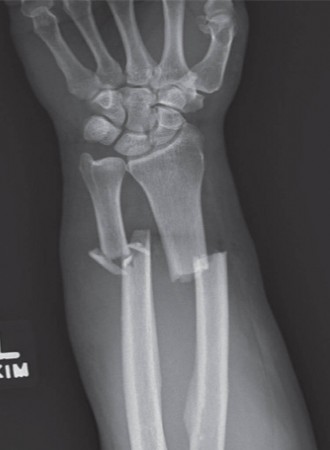
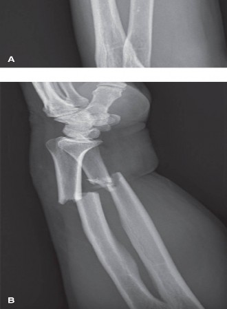
Clinical & Radiographic Imaging