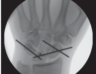
This is a lateral radiograph of the wrist that shows a perilunate dislocation. The dorsal view shows a dorsal dislocation of the capitate and distal carpal row. The lunate remains in the lunate fossa of the radius due to strong ligamentous attachments from the short and long radiolunate ligaments. The PA view would reveal gingival line disruption with capitate overlapping the lunate. There are no obvious fractures, but closely examine for any of the more commonly associated bony injuries, including fractures to the radial styloid, scaphoid, capitate, trapezoid, hamate, and ulnar styloid.
**Detailed Explanation:**
This radiograph shows a perilunate dislocation with dorsal dislocation of the capitate and distal carpal row while the lunate remains in the lunate fossa of the radius due to ligamentous attachments. The PA view would reveal gingival line disruption with capitate overlapping the lunate. Fractures to the radial styloid, scaphoid, capitate, trapezoid, hamate, and ulnar styloid are commonly associated with this injury and should be checked closely.
2. How many Gilula lines are there? Show Answer
There are three in total: the first line follows a smooth curve outlining the proximal convex surfaces of the scaphoid, lunate, and triquetrum; the second line traces the distal concave surfaces of the same bones; and the third line follows the proximal curvatures of the capitate and hamate.
Show Explanation
- The first line follows a smooth curve outlining the proximal convex surfaces of the scaphoid, lunate, and triquetrum.
- The second line traces the distal concave surfaces of the same bones.
- The third line follows the proximal curvatures of the capitate and hamate.
Click the image to enlarge.
3. How would you manage this injury in the emergency department? Show Answer
This is a potentially high-energy injury, and the patient needs concurrent assessment and treatment according to ATLS guidelines. For the affected limb, I would perform a circumferential examination to ensure it is a closed injury and assess the neurovascular status of the hand, particularly the median nerve. I would provide the patient with suitable analgesia and arrange for a closed reduction in the emergency department, utilising the Tavernier’s manoeuvre (traction, wrist extension with direct thumb pressure on the lunate, followed by wrist flexion) and place the patient in a plaster of Paris below elbow backslab. I would assess for congruent reduction with AP and lateral radiographs before discussing definitive operative management with the patient.
Show Explanation
ATLS guidelines should be followed during the management of this potentially high-energy injury. A circumferential examination should be conducted to determine if it is a closed injury, while a neurovascular status assessment should be conducted to examine the affected hand for the median nerve injury. Suitable analgesia is administered before a reduction of the closed injury using Tavernier's manoeuvre (traction, wrist extension, direct thumb pressure on the lunate, and wrist flexion). A plaster of Paris below elbow backslab is applied, and radiographs are taken to assess for congruent reduction before discussing definitive operative management with the patient.
4. How would you classify this injury? Show Answer
There are two broad classifications for this injury: greater arc and lesser arc injuries. This injury would be classified as a lesser arc, as it is purely ligamentous. There are typically four stages of lesser arc injuries, with this particular case being classified as a Mayfield stage III perilunate dislocation. Stage I involves the failure of the scapholunate ligament. Stage II involves stage I and lunocapitate joint disruption. Stage III involves stage I, II, and perillunate dislocation. Stage IV involves lunate dislocation (usually volar through the space of Poirier, which has a high rate of carpal tunnel syndrome).
Show Explanation
Wrist perilunate dislocations can be classified into two categories: greater arc injuries and lesser arc injuries. This injury is classified as a lesser arc injury as it is purely ligamentous. Mayfield devised a four-stage system that classifies these injuries. Disaster begins with the failure of the scapholunate ligament (stage I). The following stage, stage II, is the addition of lunocapitate joint disruption. The third stage, stage III, involves stage I, II, and perilunate dislocation. Finally, stage IV is lunate dislocation (usually volar through the space of Poirier, which has a high rate of carpal tunnel syndrome).
