العربية

Pediatric Orthopaedic B Review | Dr Hutaif Pediatric Or -...

Updated: Feb 2026 75 Views
Pediatric Orthopaedic B Review | Dr Hutaif Pediatric Or -...
Score: 0%

Orthopedics Online MCQs

QUESTION 1
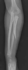
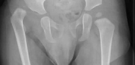
A 26-month-old boy with a displaced spiral mid-diaphyseal femur fracture with
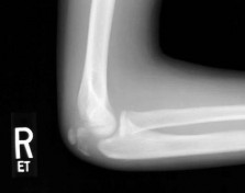
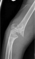
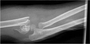
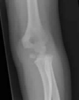
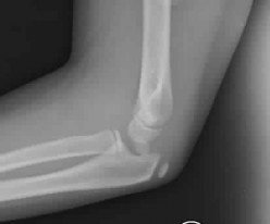
An 11-year-old boy reports the acute onset of elbow pain and swelling after pushing his brother. The patient's mother and a younger sibling have experienced numerous fractures. You note that the patient and his mother have blue sclera and normal-appearing teeth. A radiograph of the elbow is shown in Figure 60. This patient's disorder is most likely the result of ](http://www.orthobullets.com/pediatrics/4102/osteogenesis-imperfecta)Review Topic


1
parental abuse.
2
abnormal osteoclast function.
3
defective N-Ac-Gal-6 sulfate sulfatase enzyme.
4
a quantitative defect of type I collagen synthesis.
5
vitamin D deficiency. Osteogenesis imperfecta (OI) is a genetically determined disorder of type I collagen synthesis characterized by bone fragility. This patient sustained a displaced fracture of the olecranon apophysis after relatively minor trauma. Physical examination reveals distinctly blue sclera. His mother and younger sibling have experienced numerous fractures suggesting a family history of bone fragility. The patient's history, clinical features, and family history are consistent with a diagnosis of Sillence type I-A OI. Type I OI is the mildest and most common form. Inheritance is autosomal dominant. Type I is subclassified into the A type (absence of dentinogenesis imperfecta) and B type (presence of dentinogenesis imperfecta). These individuals have blue sclerae, and although the initial fracture usually occurs in the preschool years, it may occur at any age. Furthermore, olecranon apophyseal fractures that occur after relatively minor trauma have been associated with type I OI. Cells from individuals with type I OI largely demonstrate a quantitative defect of type I collagen; they synthesize and secrete about half the normal amount of type I procollagen. In this patient, there are no indications that the child has been subjected to abuse. Radiographs of the elbow show no evidence of osteopetrosis (due to abnormal osteoclast function) or rickets (due to a deficiency of vitamin D). Morquio syndrome (characterized by a defect of the enzyme N-Ac-Gal-6 sulfate sulfatase) is not associated with blue sclera. Which gene correlates with severity of disease in spinal muscular atrophy (SMA)? ](http://www.orthobullets.com/pediatrics/4091/spinal-muscular-atrophy)Review Topic
Dr. Mohammed Hutaif
Written & Medically Reviewed by
Consultant Orthopedic & Spine Surgeon