A 45-year-old male is taken to the emergency room after sustaining a motor vehicle accident. An Orthopaedic consult is placed after a unexpected finding is seen on the X-ray of his left hip. The X-rays is shown is Figure 8–40A and an MRI is shown is Figure 8–40B.
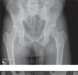
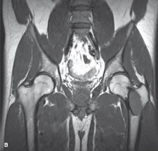
Figure 8–40 A–B
What is the most likely diagnosis?
- Osteosarcoma
- Chronic osteomyelitis
- Aneurysmal bone cyst
- Fibrous dysplasia
Discussion
The correct answer is (D). Fibrous dysplasia is characterized by a fibro-osseous tissue within the bone and is often asymptomatic and found incidentally. Lesions can
be expansile with cortical thinning and a sclerotic rim, around central lucent lesions within the medullary canal. When it occurs in the proximal femur, a coxa vara deformity can result, termed the “shepherd’s crook” deformity.
The biopsy of this lesion is shown inFigure 8–41. What of the following histologic features is characteristic of this lesion? 1. “Alphabet soup” pattern of bony trabeculae
2. Chondroid, myxoid, and fibrous areas
3. Bland cartilage
4. Areas of aneurysmal degeneration
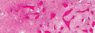
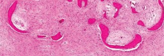
Figure 8–41
Discussion
The correct answer is (A). Histologically, fibrous dysplasia appears as poorly mineralized immature fibrous tissue surrounding islands of irregular trabeculae of woven bone, termed “Chinese letters” or “alphabet soup.” These lesions can be associated with secondary aneurysmal bone cysts, so sometimes cavernous blood-filled spaces are seen on histology, but this is not a characteristic feature and cannot be used alone to make the diagnosis.
He has no symptoms in his left hip. What is the most appropriate management at this time?
- Observation
- Curettage and bone graft using cortical allograft
- Curettage and bone graft using cancellous allograft
- Arthroplasty
Discussion
The correct answer is (A). Asymptomatic patients may be observed. Surgical indications include painful lesions, impending/actual pathologic fracture, severe deformity, and neurologic compromise. When curettage and bone grafting is performed, cortical allograft should be used. Internal fixation is usually recommended to achieve adequate stabilization and pain control. Objectives: Did you learn...?
To recognize fibrous dysplasia?
To understand the treatment options for impending fracture?
