Score: 0%
ORTHOPEDIC MCQS ONLINE 014 PATHOLOGY
QUESTION 1
ORTHOPEDIC MCQS ONLINE 014 PATHOLOGY
..External beam radiotherapy


..External beam radiotherapy
1
should include the instrumented femur and periacetabular area.
2
should include the femur only.
3
should include the acetabulum only.
4
is contraindicated for this patient.
..: 3- CT scan of the chest, abdomen, and pelvis. PREFERRED RESPONSE2…: 1- Metastatic adenocarcinoma
…: 4- Complex total hip arthroplasty
PREFERRED RESPONSE4 …: 1- should include the instrumented femur and periacetabular area.
..Li-Fraumeni syndrome (LFS) is associated with
1) multiple hemangiomas.
2) multiple hereditary osteochondromatosis.
3) soft-tissue sarcomas.
4) neurofibromatosis.
- soft-tissue sarcomas.
..A 60-year-old woman has a proximal femur fracture. A permeative, lytic defect is recognized at the fracture site. Appropriate imaging studies are performed and show no other lesions. What is the next treatment step?
1) Cephalomedullary nail
2) Standard antegrade intramedullary nail
3) Resection and arthroplasty reconstruction
4) Open biopsy
- Open biopsy
CLINICAL SITUATION FOR QUESTIONS 7 THROUGH 9
…Based on the images and histopathology, how is this patient best treated?
1) Chemotherapy and external beam radiotherapy
2) Resection
3) Resection and chemotherapy
4) External beam radiation alone
…: 3- Chordoma PREFERRED RESPONSE14…: 2- Resection
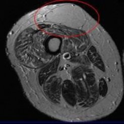
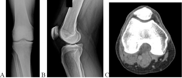
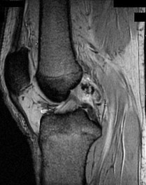
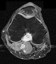
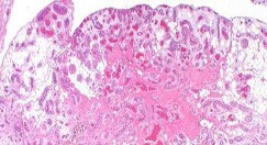
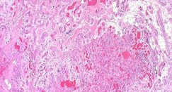
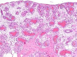
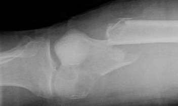
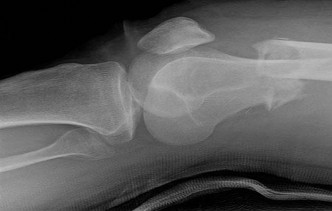
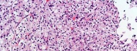
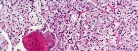
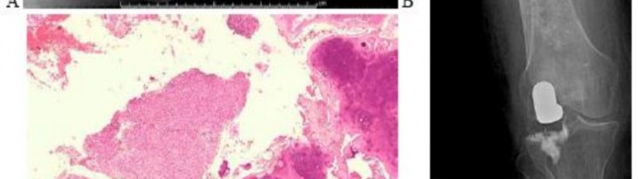
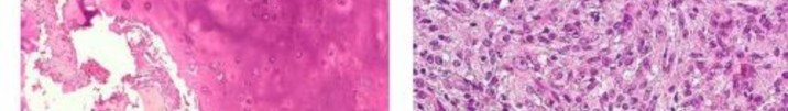
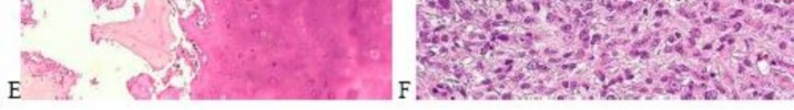
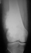
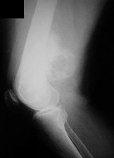
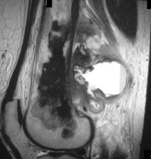
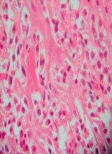
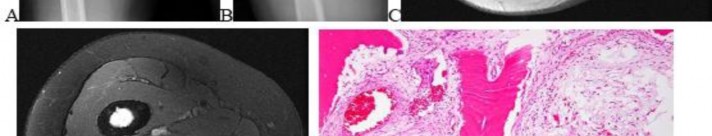
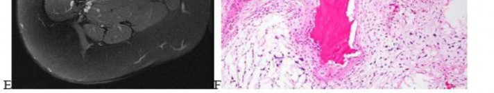
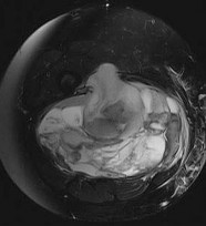
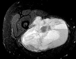
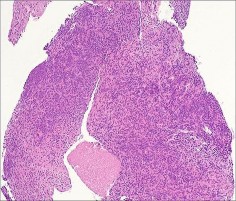
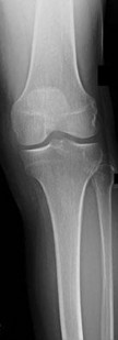
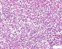
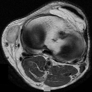
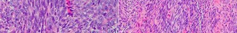
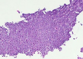
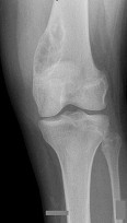
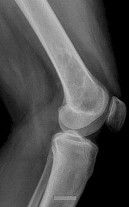
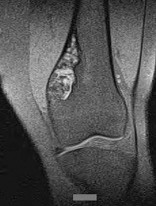
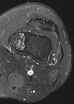
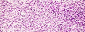
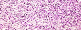
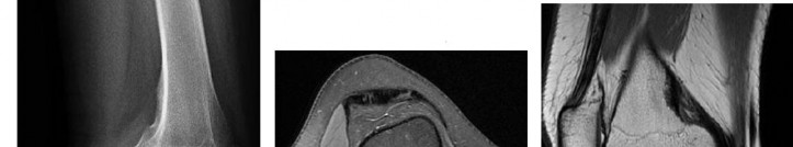
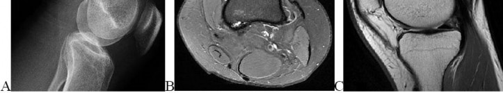
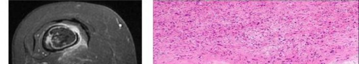
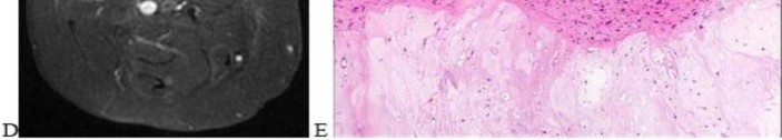
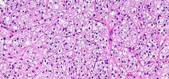
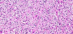
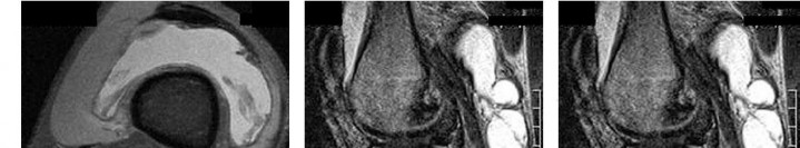
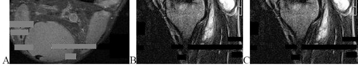
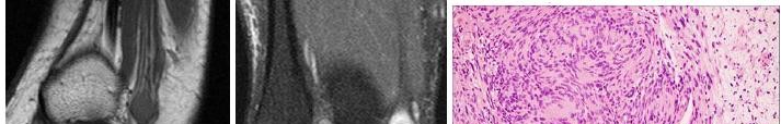
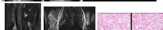
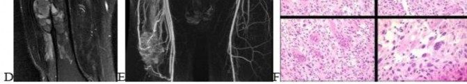
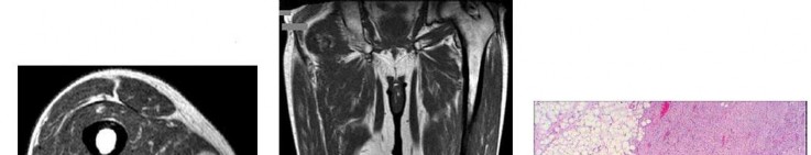
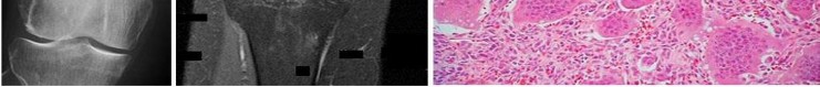
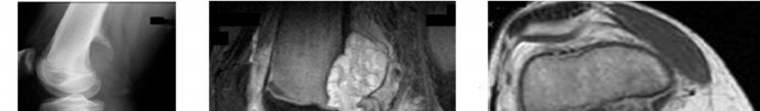
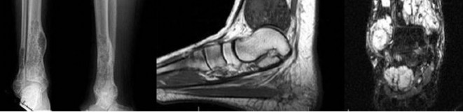
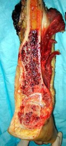
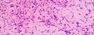
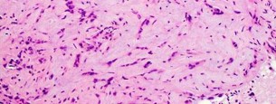
…A 56-year-old podiatrist with a negative past medical history had anterior knee pain after an injury. His radiographs, CT scan, and T1-weighted sagittal and fat-saturated axial MR images are shown in Figures 15a through 15e, respectively. After arthroscopic partial medial menisectomy, the patient was turned to the prone position and an open posterior arthrotomy and excision was performed. Low-power and high-power hematoxylin and eosin stained histologic specimens are shown in Figures 15f and 15g, respectively. Based on the history, radiographs, CT scan, MRI scans, and histologic findings, what is the most likely diagnosis?
1) Localized pigmented villonodular synovitis (PVNS)
2) Biphasic synovial sarcoma
3) Nodular fasciitis
4) Synovial hemangioma
- Synovial hemangioma
CLINICAL SITUATION FOR QUESTIONS 16 THROUGH 19
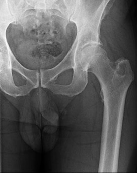
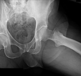
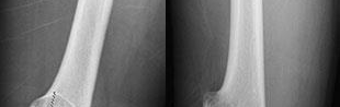
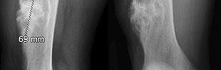
Figures 16a and 16b are the right femur radiographs of a 59-year-old man who has severe pain in his right distal thigh and knee with no significant trauma and an inability to bear weight. Blood tests demonstrate anemia, serum protein electrophoresis/urine protein electrophoresis findings are negative, and electrolyte levels are within defined limits.
..Approximately what percentage of the time does an unknown primary cancer get identified as part of a full metastatic work-up that includes radiographs; blood tests; a CT scan of the chest, abdomen and pelvis; whole-body bone scan; and biopsy of the metastatic focus?
1) 45%
2) 65%
3) 85%
4) 100%
..: 3- Biopsy of the fracture site
PREFERRED RESPONSE 18..: 3- Distal femoral resection with megaprosthesis PREFERRED RESPONSE 19..: 3- 85%...
CLINICAL SITUATION FOR QUESTIONS 20 THROUGH 23
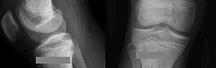
Figures 20a and 20b are the radiographs of an 83-year-old active, independent, and healthy woman who has experienced 2 months of right lower thigh and knee pain. Her pain increased progressively over the course of several weeks. While exiting a car she “bumped” her knee against the door, felt a “crack,” and developed excruciating pain. She could no longer ambulate and was brought to the hospital.
..What is the most likely site of metastatic disease in patients with this diagnosis?
1) Liver
2) Lungs
3) Brain
4) Kidneys
-: 4- Enchondroma
PREFERRED RESPONSE21-..: 4- Dedifferentiated chondrosarcomas
PREFERRED RESPONSE22-..: 3- Above-the-knee amputation with wide surgical margin PREFERRED RESPONSE23-..: 2- Lungs
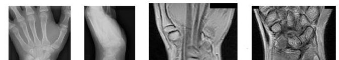
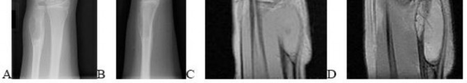
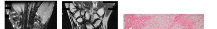
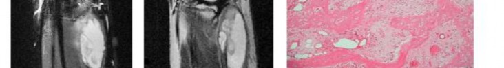
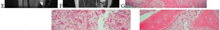
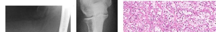
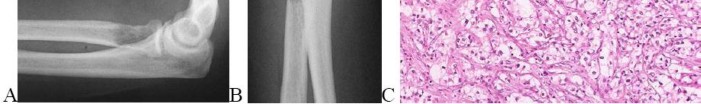
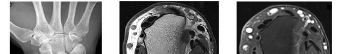
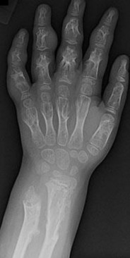
..A 14-year-old boy has had wrist pain for 3 weeks. Radiographs are shown in Figures 24a and 24b. His MRI scans are shown in Figures 24c through 24f. Representative histology is shown in Figures 24g through 24i. The most likely diagnosis is
1) aneurysmal bone cyst.
2) fibrous dysplasia.
3) giant-cell tumor.
4) osteoblastoma.
- osteoblastoma.
RESPONSES FOR QUESTIONS 25 THROUGH 29
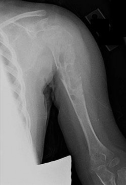
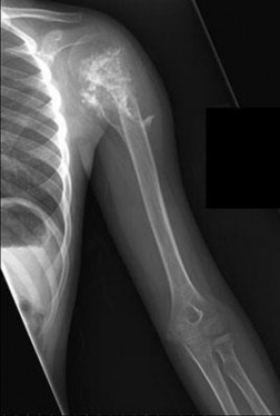
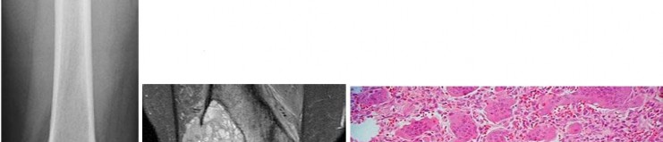
..Figure 30a is the radiograph taken in the emergency department of a 20-year-old man with pain and swelling in his right-dominant arm. His MRI scan is seen in Figure 30b, and his histopathology is shown in Figure 30c. What is the most likely diagnosis?
1) Ewing sarcoma
2) Langerhans cell histiocytosis
3) Osteosarcoma
4) Osteomyelitis
- Ewing sarcoma
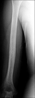
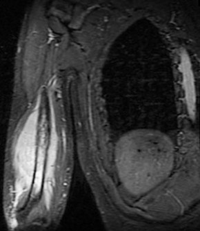
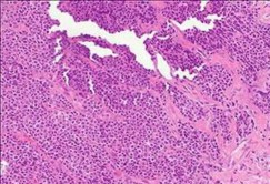
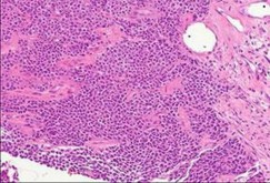
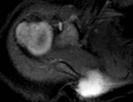
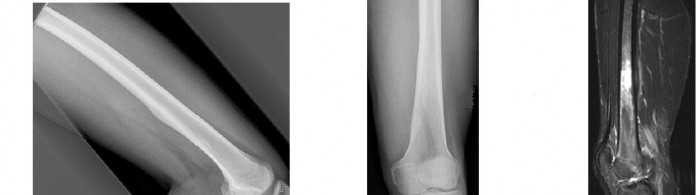
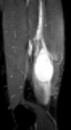
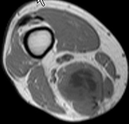
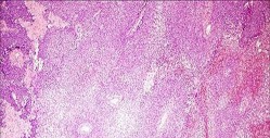
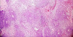
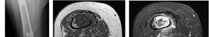
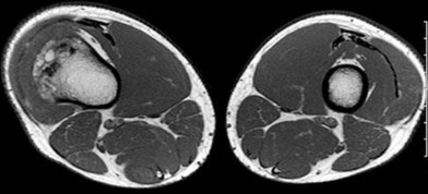
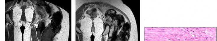
..An 18-year-old woman has had a right thigh mass for “a long time.” She has found it increasingly difficult to ambulate. Her medical history is unremarkable, with the exception of hypertension. On clinical examination, a fullness is appreciated in the popliteal fossa. The neurovascular status of the distal extremity is intact. Anteroposterior and lateral radiographs are shown in Figures 31a and 31b. A lateral T2-weighted MRI scan is shown in Figure 31c. The histology is seen in Figure 31d. What is the most likely diagnosis?
1) Aneurysmal bone cyst
2) Osteosarcoma
3) Periosteal lipoma
4) Parosteal osteosarcoma
- Osteosarcoma
CLINICAL SITUATION FOR QUESTIONS 32 THROUGH 36
A 68-year-old woman is referred for left thigh pain. Her medical history includes Hypertension, diabetes, and adenocarcinoma of the breast treated with surgery, chemotherapy, and radiation 3 years ago. She currently is on aromatase therapy. She is unable to ambulate secondary to pain, is limited to a walker, and requires narcotic medications. She has no other pain but agrees to your recommendation that she urgently be sent to the hospital.
..Which intervention should be added to this patient’s care to best prevent future skeletally related events (SRE)?
1) Inferior vena cava (IVC) filter placement
2) Bisphosphonates
3) External beam radiation
4) Tc-99 whole-body bone scan at regular intervals
…: 3- Further imaging PREFERRED RESPONSE 33-..: 3- Location of the lesion PREFERRED RESPONSE 34-…: 2- 15%
PREFERRED RESPONSE 35…..:4- Lung carcinoma, breast carcinoma, multiple myeloma PREFERRED RESPONSE 36-…: 2- Bisphosphonates
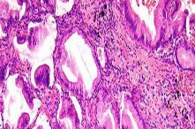
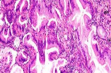
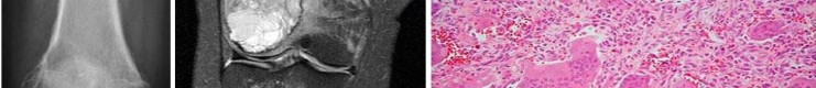
..Figures 37a and 37b are the lateral and anteroposterior (AP) radiographs of a 60-year-old man with a remote history of renal cell cancer. A needle biopsy of the lesion is shown in Figure 37c. The bone destruction that occurs in this process is a result of
1) tumor cells.
2) cytokines secreted by the tumor.
3) host bone osteoblasts.
4) osteoprotegerin.
.
- cytokines secreted by the tumor
..Figures 38a and 38b are the histopathology of an otherwise healthy 31-year-old man who had a growing mass excised from his forearm with local anesthetic and no preoperative imaging. The mass was documented to be subfascial and larger than 5 cm.
What is the best local treatment option?
1) Observation
2) Radiation only
3) Chemotherapy only
4) Re-excision and radiation
- Re-excision and radiation
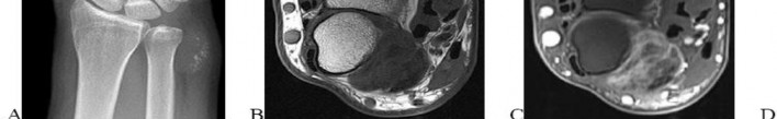
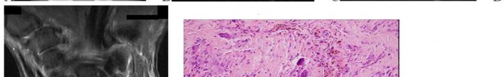
…Figure 39a is the anteroposterior radiograph of a 51-year-old man who has had a painless soft-tissue mass on his left wrist for 2 months. MR sequences are shown in Figures 39b through 39d. A biopsy was performed and shown in a low-power hematoxylin and eosin photomicrograph in Figure 39e. The most appropriate treatment for this lesion is
1) a diet that reduces uric acid production.
2) wide local resection followed by radiotherapy.
3) marginal excision.
4) observation until the mineralization matures, and then excision and radiotherapy to prevent recurrence.
- marginal excision.
CLINICAL SITUATION FOR QUESTIONS 40 THROUGH 43
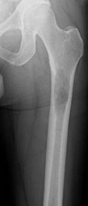
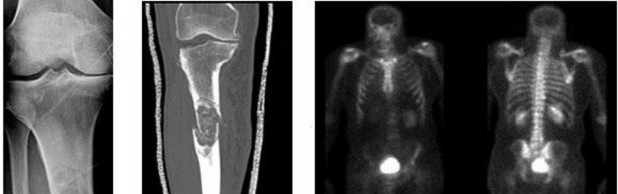
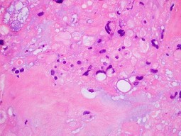
Figures 40a through 40c are the radiograph, bone scan, and histology of a 68-year-old man who has had 3 months of pain in his left thigh with weight bearing. He has no history of cancer and no illnesses.
..The orthopaedic surgeon obtains tissue with the histology shown in Figure 40c. Treatment should consist of
1) surgical stabilization.
2) surgical stabilization and radiation.
3) excision and endoprosthesis.
4) radiation.
- …..: 1- malignant.
PREFERRED RESPONSE 41..,,,,,,,: 1- CT scan of the chest, abdomen and pelvis. PREFERRED RESPONSE 42………: 2- biopsy.
PREFERRED RESPONSE 43……….: 2- surgical stabilization and radiation.
..First-line treatment recommendations include
1) synovectomy.
2) arthrocentesis, compressive wrap, and rest.
3) en bloc resection.
4) intra-articular radioactive nucleotide injection.
- pigmented villonodular synovitis (PVNS). PREFERRED RESPONSE: 1- synovectomy.
RESPONSES FOR QUESTIONS 47 THROUGH 52
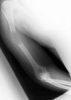
…Figure 53 is the emergency department radiograph of a 7-year-old boy who has pain and is unwilling to use his right arm after a fall on the playground. What is the most appropriate initial treatment?
1) Nonsurgical treatment of the fracture
2) Aspiration and injection with methylprednisolone
3) Curettage and augmentation with bone cement and internal fixation
4) Further imaging and biopsy
- Nonsurgical treatment of the fracture
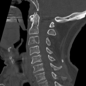
..Figure 54 is the CT scan of a 70-year-old man with progressive neck pain; there is no history of trauma, and examination is notable only for mildly decreased cervical range of motion. He is neurologically intact. He has monoclonal gammopathy of undetermined significance that has been stable for many years. Current serum protein electrophoresis is unchanged. History and examination reveal no other causes for his pain. What is the next step in clinical management?
1) Corpectomy and anterior fusion
2) Radiation therapy followed by multiple myeloma protocol chemotherapy
3) CT-guided biopsy
4) CT scan of the chest, abdomen, and pelvis
- CT scan of the chest, abdomen, and pelvis
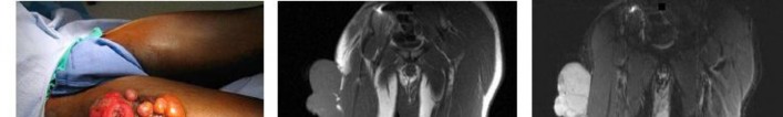
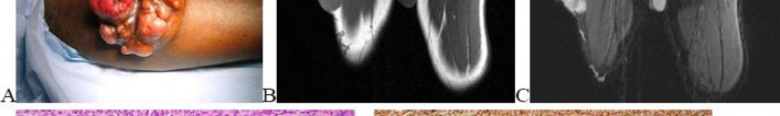
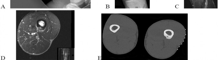
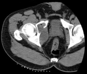
…A 27-year-old incarcerated man was found to have a fungating mass on his anterolateral right proximal thigh. A clinical photograph is shown in Figure 55a. T1- and T2-weighted coronal MRI scans are shown in Figures 55b and 55c. The hematoxylin and eosin and CD34 stained histology are shown in Figures 55d and 55e. What is the most likely diagnosis?
1) Squamous cell carcinoma
2) Melanoma
3) Dermatofibrosarcoma protuberans (DFSP)
4) Desmoid tumor
- Dermatofibrosarcoma protuberans (DFSP)
..Figures 56a and 56b are the axial short tau inversion recovery and T1 with contrast images of a 7-month-old infant who is found to have a right scapular soft-tissue mass. On examination, the mass is hard. A biopsy was performed and is shown in Figure 56c (hematoxylin and eosin, 400x). What is the optimal treatment for this patient?
1) Intralesional excision 2- Marginal excision
2) Wide excision 4- Observation
- Wide excision
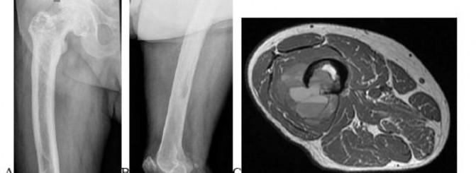
…Figures 57a through 57d show the radiographs and T1- and T2-weighted MRI scan sequences of the proximal femur of a 60-year-old man with progressive thigh pain. A review of radiographs taken 3 years ago reveals that the lesion is new. The biopsy specimen is shown in Figure 57e; staging studies show no other lesions, and local imaging confirms the process is confined to bone (no soft-tissue mass). The next treatment step should include
1) hip disarticulation.
2) radiotherapy.
3) ifosfamide-based chemotherapy.
4) wide resection and reconstruction.
- wide resection and reconstruction.
…The characteristic translocation and genes involved in extraskeletal myxoid chondrosarcoma is
1) t(11;12) EWS;FLI1
2) t(12;16) TLS;CHOP.
3) t(9;22)EWS;CHN.
4) t(9;22) BCR-ABL.
.
- t(9;22) EWS;CHN
.Figures 59a and 59b are the axial T2 and T1 with contrast MRI scans of a 32-year-old woman who has a 10-year history of pain and a 1-year history of progressive swelling in her right leg. The histopathology is shown in Figure 59c. A radiograph of her leg showed no mineralizations or osseous erosions. The chromosomal abnormality that is associated with this disease is
1) t(11;22).
2) t(2;13).
3) t(X;18).
4) t(12;16).
- t(X;18).
CLINICAL SITUATION FOR QUESTIONS 60 THROUGH 63
A 45-year-old woman has an enlarging buttock mass. The mass is 12 cm and nonpainful. The patient first noticed it about 6 months after she had a low-impact fall. The general surgeon evaluating the patient felt this mass could be either a lipoma or a hematoma. The patient underwent a surgical procedure to remove the mass.
..What is the most common detrimental impact of an unplanned excision of a high-grade soft-tissue sarcoma?
1) Decreased mortality
2) Decreased recurrence
3) Increased wound complications
4) Increased functional outcome
-,…: 2- Imaging studies (MRI scan or CT scan) PREFERRED RESPONSE 61….: 1- Meticulous hemostasis and closure PREFERRED RESPONSE 62…..: 3- No imaging was obtained before surgery. PREFERRED RESPONSE 63…..: 3- Increased wound complications
CLINICAL SITUATION FOR QUESTIONS 64 THROUGH 66
Figures 64a through 64c are the radiograph, MRI scan, and histology of a 53-year-old man with medial knee pain and swelling below the knee.
..Histology of the lesion is shown in Figure 64c. The best next treatment step is
1) radiation.
2) radiation and surgery.
3) chemotherapy.
4) observation.
- Translocation x;18 PREFERRED RESPONSE: 4- Biopsy
PREFERRED RESPONSE: 2- radiation and surgery.
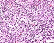
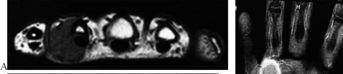
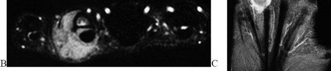
..A 26-year-old woman has had a slow-growing and painful mass at the base of her ring finger for several months. Radiographs of the affected digit show no mineralization or erosions of the underlying bone. An axial T1 MRI scan is shown in Figure 67a, and a corresponding short tau inversion recovery image is shown in Figure 67b. A coronal T1 MRI scan with contrast is shown in Figure 67c. The best next treatment step is
1) referral to a sarcoma center.
2) observation.
3) excisional biopsy.
4) marginal excision.
- referral to a sarcoma center.
..What is the most specific immunohistochemistry staining pattern that confirms the diagnosis of desmoid tumor?
1) Membranous beta-catenin staining
2) Nuclear beta-catenin staining
3) Nuclear SMAD4 staining
4) Vimentin positivity
- Nuclear beta-catenin staining
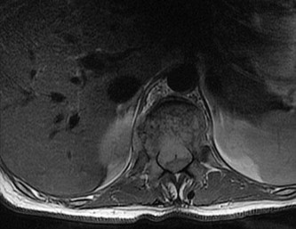
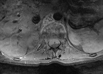
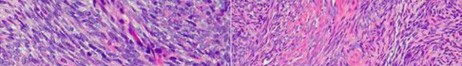
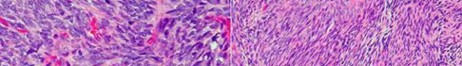
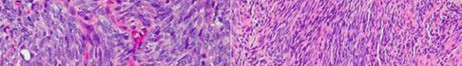
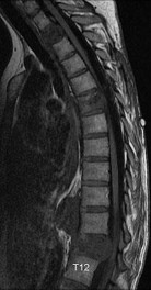
…A 30-year-old woman has progressive gait instability, back pain, and urinary retention. Figures 69a and 69b show the axial T2 and postcontrast MRI scans taken at the level of T11, and Figure 69c shows the sagittal T1-weighted image. Representative histology is shown in Figure 69d. The patient remains ambulatory, but symptoms have
progressed during the last week and she is beginning to feel weakness in her legs. Examination is notable for decreased rectal tone, lower-extremity hyperreflexia and clonus, and 4/5 motor strength throughout the lower extremities. What is the most appropriate treatment recommendation for this patient?
1) Margin-free en bloc spondylectomy of T11
2) Radiation therapy
3) Radiation therapy followed by anterior corpectomy and fusion
4) Transpedicular decompression and posterior stabilization followed by radiation therapy
- Transpedicular decompression and posterior stabilization followed by radiation therapy
…Giant-cell tumor of bone usually involves the epiphysis of long bones. What is the next most common type of tumor involving this anatomical location?
1) Conventional chondrosarcoma
2) Aneurysmal bone cyst
3) Chondroblastoma
4) Osteoblastoma
- Chondroblastoma
CLINICAL SITUATION FOR QUESTIONS 71 THROUGH 73
Figures 71a through 71e are the radiographs, MRI scan, and CT scans of a 14-year-old-boy who has cyclical pain in his thigh. His symptoms began approximately 6 months ago. He complains of increased pain when he runs and also of pain that wakes him at night. This pain is relieved by nonsteroidal anti-inflammatory drugs (NSAIDs).
…What is the etiology of the pain associated with this lesion?
1) Prostaglandin production
2) Gram-positive cocci
3) Osteoclast activation
4) Loss of structural integrity of the bone
- Osteoid osteoma PREFERRED RESPONSE: 2- radiofrequency ablation. PREFERRED RESPONSE: 1- Prostaglandin production
...Figure 74 is the radiograph of an 11-year-old boy with pain in his left arm. Prognosis is most influenced by
1) stage at presentation.
2) grade at presentation.
3) response to neoadjuvant chemotherapy.
4) histologic subtype.
- stage at presentation.
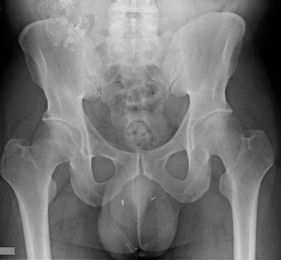
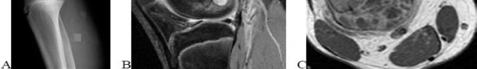
..Figures 75a through 75d are the radiograph, CT scan, bone scan, and biopsy of a 45-year-old man who has had a several-month history of progressive pain in his right hip and groin region. Based on these images and histology, what is the most appropriate treatment?
1) Wide resection
2) Curetting and bone grafting
3) Percutaneous cementation and radiotherapy
4) Chemotherapy and radiotherapy
- Wide resection
..Figure 76 is the radiograph of a 77-year-old patient with a history of myeloma who has had severe arm pain after opening a jar. Pain was present for 3 months prior to injury. The most biomechanically stable construct for this fracture is
1) intramedullary nailing (IMN).
2) IMN and cement.
3) plate.
4) plate and cement.
- plate and cement.
CLINICAL SITUATION FOR QUESTIONS 77 THROUGH 79
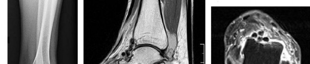
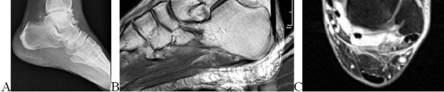
Figures 77a through 77c are the radiograph and MRI scans of a 45-year-old woman who enjoys dancing and has had left ankle pain for approximately 1 month with minimal trauma. She has slightly limited ankle dorsiflexion with a mechanical block with pain.
..The patient undergoes recommended surgery and minimal joint damage is found. How should she be counseled regarding her future prognosis?
1) High risk for local recurrence, moderate risk for metastases
2) High risk for local recurrence, no risk for metastases
3) Low risk for local recurrence, moderate risk for metastases
4) Low risk for local recurrence, no risk for metastases
- Synovial metaplasia
PREFERRED RESPONSE: 2- TA, EHL, anterior tibial artery, deep peroneal nerve, EDL PREFERRED RESPONSE: 4- Low risk for local recurrence, no risk for metastases
..Figures 80a through 80d are the radiographs and MRI scans of a 16-year-old girl who requires crutches because she is unable to bear weight on her right lower extremity. The pain has progressed over 2 months despite nonsurgical treatment.
Treatment at this point should include
1) continued observation.
2) aspiration and injection with methylprednisolone.
3) curettage and grafting.
4) wide margin resection.
- curettage and grafting.
..A previously healthy 60-year-old woman has a T5 pathologic compression fracture secondary to metastatic renal cell carcinoma. She has myelopathy with severe pain upon sitting from a supine position. The adjacent thoracic levels are unaffected. On MRI scan, there is minimal space available for the cord because of epidural involvement. What is the optimal treatment method at this time?
1) Surgical decompression and instrumented fusion
2) Surgical decompression without fusion
3) Image-guided intensity-modulated radiation therapy
4) Chemotherapy
- Surgical decompression and instrumented fusion
..Figures 82a and 82b are the MRI scans consisting of a T2 coronal sequence and axial T1 pulse sequence of a 38-year-old man who has had right thigh pain and a mass for 4 months since he pulled his hamstring. The presumed diagnosis considering his clinical history and evaluation of the MRI scan was hematoma and the mass was evacuated. The histology is shown in Figures 82c and 82d. Next treatment steps should include
1) physical therapy to accelerate healing and improve function.
2) a complete hematologic work-up to evaluate a bleeding disorder.
3) tumor bed excision and radiation to reduce local recurrence.
4) radiotherapy to complete definitive treatment of this problem.
- tumor bed excision and radiation to reduce local recurrence.
….Figures 83a through 83c are the radiograph and MRI scans of a 16-year-old girl who had posterior knee pain after a dance recital 3 weeks ago; the pain resolved 1 week ago with ibuprofen use. What is the appropriate treatment for this patient?
1) Biopsy and resection of lesion
2) Observation and serial radiographs
3) Tc-99 whole-body bone scan and fine-cut CT scan
4) Evaluation by a pediatric oncologist
- Observation and serial radiographs
CLINICAL SITUATION FOR QUESTIONS 84 THROUGH 86
Figures 84a and 84b are the CT and MRI scans of a 17-year-old girl with a painful lumbosacral scoliosis that has been present for 12 months. Examination is notable only for pain over the left sacral region and a postural scoliosis leaning away from this side.
..With treatment, the spinal deformity is expected to
1) spontaneously resolve.
2) remain stable and nonprogressive.
3) respond in proportion to the family’s compliance with brace treatment.
4) resolve in the coronal plane and progress in the sagittal plane.
- is likely to respond to percutaneous intervention.
PREFERRED RESPONSE: 1- uses nonsteroidal anti-inflammatory or aspirin medications. PREFERRED RESPONSE: 1- spontaneously resolve.
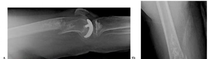
..Figures 87a through 87e are the radiograph, MRI scans, and biopsy specimen of an 83-year-old woman who is experiencing pain in her distal thigh with activity and at night. She has undergone total hip arthroplasty for hip osteoarthritis. The most appropriate treatment is
1) external beam radiation.
2) curetting and cementation.
3) radiofrequency ablation.
4) wide local resection.
- wide local resection.
CLINICAL SITUATION FOR QUESTIONS 88 through 92
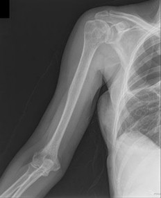
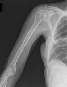
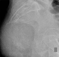
Figures 88a and 88b are the radiographs of a 70-year-old woman with a remote history of localized renal cell carcinoma. She has insidious onset of right shoulder pain that worsens with any activity and at night. She appears otherwise healthy.
..Oncologic outcome for this patient
1) is influenced by extraosseous tumor extension.
2) is more favorable if the lesion is solitary.
3) involves a predictable rapid demise.
4) hinges on the presence or absence of gene amplification.
- CT scan of the chest, abdomen, and pelvis.
PREFERRED RES: 3- Should be performed if the lesion is solitary, but not necessarily if multifocal PREFERRED RESPONSE: 4- resection and reconstruction.
PREFERRED RESPONSE: 3- inhibits vascular endothelial growth factor (VEGF) pathways. PREFERRED RESPONSE: 2- is more favorable if the lesion is solitary.
..Figures 93a and 93b are the MRI scans of a 24-year-old man with painless, persistent swelling in his left knee without any trauma. What is the best next treatment step?
1) Arthroscopic anterior synovectomy and posterior open resection
2) Arthroscopic anterior synovectomy only
3) Radiation therapy and wide excision
4) Observation
- Arthroscopic anterior synovectomy and posterior open resection
..What biopsy technique for a posterior thigh sarcoma is associated with the highest risk for adverse outcome?
1) Transverse incision open biopsy
2) Core needle biopsy
3) Fine-needle aspirate
4) Longitudinal incision open biopsy
- Transverse incision open biopsy
..A 60-year-old woman with a history of breast cancer has a rapidly enlarging arm mass. The lesion is situated outside of the prior irradiation field, but within an area of heavy lymphedema involvement. Needle biopsy reveals a high-grade sarcoma. What is the most likely diagnosis?
1) Lymphangiosarcoma
2) Acral myxoinflammatory fibroblastic sarcoma
3) Hemangioendothelioma
4) Hemangiopericytoma
- Lymphangiosarcoma
RESPONSES FOR QUESTIONS 96 THROUGH 100
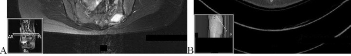
..Figures 106a and 106b are the T1 sagittal and T2 coronal images of a 41-year-old woman who has been experiencing electric shock-type radiating pain over her right ankle for 4½ years. Her biopsy specimen is shown in Figure 106c. What is the best next treatment step?
1) Marginal excision
2) Wide excision
3) Wide excision and radiation
4) Observation
- Marginal excision
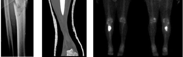
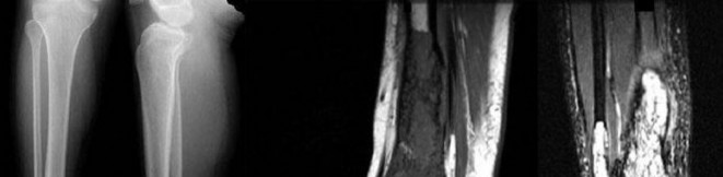
..Figures 107a through 107c are the radiograph, CT, and bone scan of a 68-year-old man. While walking, he collapsed and was unable to ambulate because of pain and deformity in his right leg. What is the most appropriate next step?
1) Staging studies to assess the extent of disease
2) Immediate stabilization of the fracture with an intramedullary nail
3) Curetting and bone culture
4) Segmental resection of the tibia and allograft reconstruction
- Staging studies to assess the extent of disease
RESPONSES FOR QUESTIONS 108 THROUGH 111
..Figures 112a and 112b are the anteroposterior and lateral radiographs of a 65-year-old man who has a significant history of tobacco abuse and a 6-week history of right thigh pain. Axial and sagittal MRI scans are seen in Figures 112c and 112d. His MR angiogram is shown in Figure 112e. A biopsy of a lesion is shown in Figure 112f. What is the most likely diagnosis?
1) Secondary sarcoma in a pre-existing condition
2) Angiosarcoma
3) Metastatic lung carcinoma
4) Fibrous dysplasia
- Secondary sarcoma in a pre-existing condition
CLINICAL SITUATION FOR QUESTIONS 113 THROUGH 116
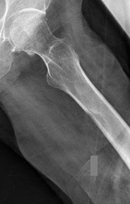
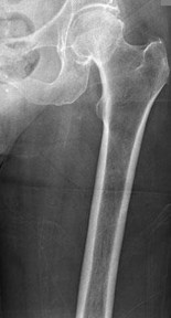
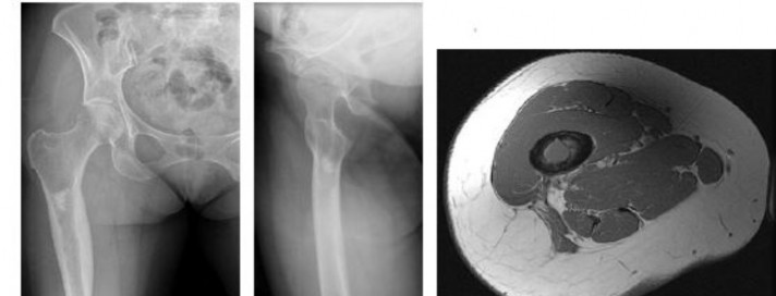
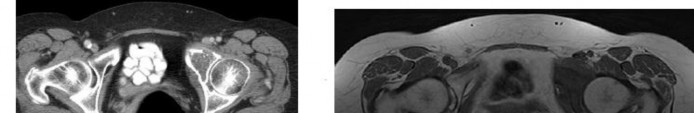
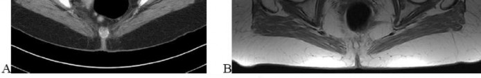
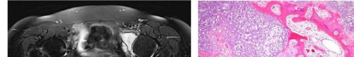
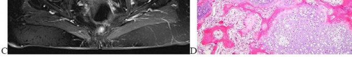
Figures 113a and 113b are the radiographs of a 68-year-old-man who has increasing pain in his left groin with weight-bearing activities and a Trendelenburg gait. Radiographs reveal a lytic lesion of the greater trochanter. An initial diagnosis of adenocarcinoma of the lung was made 1 year before this presentation. His lung cancer treatment consisted of partial lobectomy and postsurgical radiation therapy.
..Staging studies show no other lesions and surgical treatment is planned; when should a biopsy be performed?
1) Before surgery
2) Intraoperatively after instrumentation
3) After surgery (reamings/curettings sent)
4) No biopsy is needed
- Positron emission tomography (PET) scan PREFERRED RESPONSE: 3- Curettage, cementation, and internal fixation PREFERRED RESPONSE: 4- resection and prosthetic reconstruction.
PREFERRED RESPONSE: 1- Before surgery
CLINICAL SITUATION FOR QUESTIONS 117 THROUGH 120
Figures 117a through 117c are the radiographs and MRI scan of a 16-year-old boy who has had a persistent fullness in his thigh since being kicked while playing soccer 4 weeks ago. He states that initially the area was painful, but now all symptoms other than the mass have resolved.
Findings of multiple lesions in multiple skeletal sites may be associated with
1) decreased risk for malignancy.
2) a characteristic chromosomal translocation.
3) soft-tissue hemangiomas.
4) limb deformity and short stature.
- Osteochondroma
PREFERRED RESPONSE: 1- benign and simply can be observed with serial radiographs. PREFERRED RESPONSE: 4- growth beyond skeletal maturity.
PREFERRED RESPONSE: 4- limb deformity and short stature.
..Figure 121a is the axial T1 MRI scan and Figure 121b is the coronal T1 MRI scan of an 85-year-old man who has a mass in his medial thigh. The mass was present for years and recently grew. His biopsy specimen is shown in Figure 121c. What is the best treatment for this patient?
1) Chemotherapy and wide local resection
2) Wide local resection and radiotherapy
3) Marginal excision
4) Observation and reimaging in 6 months
- Wide local resection and radiotherapy
..What tumor commonly metastasizes to regional lymph nodes?
1) Fibromyxoid sarcoma
2) Epithelioid sarcoma
3) Leiomyosarcoma
4) Liposarcoma
- Epithelioid sarcoma
..A 64-year-old woman has significant right arm pain associated with a destructive proximal humeral bone lesion with an associated soft-tissue mass. Her medical history includes carcinoma of the breast treated 8 years ago with modified radical mastectomy, hormone receptor-based chemotherapy, and 45 Gy of radiation with 8 subsequent disease-free years. Biopsy reveals a high-grade osteogenic sarcoma. What factor is most likely related to her current disease?
1) Presence of metastatic disease
2) Dosage of radiation treatment
3) Type of prior surgical procedure
4) Type of chemotherapy given
- Dosage of radiation treatment
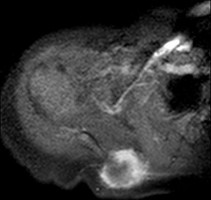
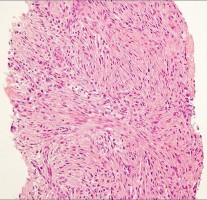
..Figures 124a and 124b are the MRI scans of a 25-year-old woman who has a painful mass in her left gluteal and thigh region. Her biopsy specimen is seen in Figure 124c. What is the most likely diagnosis?
1) Desmoid fibromatosis
2) Extraskeletal Ewing sarcoma
3) Metastatic breast cancer
4) Lymphoma
- Desmoid fibromatosis
CLINICAL SITUATION FOR QUESTIONS 125 THROUGH 128
A 45-year-old woman has increasing knee pain with activity and at rest. Her radiograph, MRI scan, and histology are shown in Figures 125a through 125c.
..The cell that directly causes osteolysis in this lesion is
1) giant cell.
2) stromal cell.
3) osteoblast.
4) fibroblast.
- benign aggressive. PREFERRED RESPONSE: 4- chest radiograph.
PREFERRED RESPONSE: 1- extended intralesional curettage. PREFERRED RESPONSE: 1- giant cell.
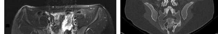
..Figures 129a through 129c are the lateral radiograph of the sacrum, axial CT scan, and a high-power view of a fine-needle biopsy of a 47-year-old man who has buttock pain and constipation. A large mass is palpable on rectal examination. The tumor cell that is the signature of this tumor is known as a(n)
1) giant cell.
2) physaliferous cell.
3) chondroblast.
4) adipocyte.
- physaliferous cell.
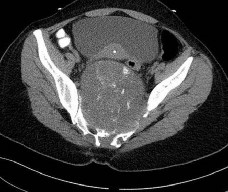
..Figures 130a through 130c show the CT scan and T1- and T2-weighted MRI scan sequences of an otherwise healthy 67-year-old woman with progressive left groin pain. Her biopsy specimen is shown in Figure 130d. Staging studies reveal no other lesions. Treatment should include
1) radiotherapy.
2) chemotherapy followed by surgical resection.
3) curettage with adjuvant treatment and grafting.
4) en bloc resection.
- en bloc resection.
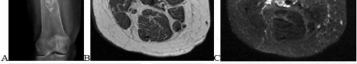
..A lateral radiograph (Figure 131a), sagittal short tau inversion recovery MRI scan (Figure 131b), and an axial T1 contrast MRI scan (Figure 131c) were performed on a 15-year-old boy who has injured his right knee twice during the last 5 months. He has a reduced range of motion of the knee and posterior thigh tenderness. A biopsy showed bland spindle cells, giant cells, and blood-filled spaces without endothelial lining. What is the most appropriate treatment?
1) Chemotherapy and wide local excision
2) Chemotherapy and radiotherapy
3) Marginal excision
4) Extended intralesional curettage
- Extended intralesional curettage
CLINICAL SITUATION FOR QUESTIONS 132 THROUGH 134
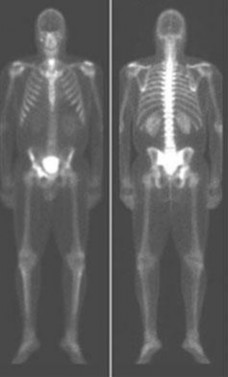
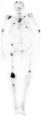
Figure 132 is the bone scan of a 73-year-old man who is referred from his family doctor with diffuse bone pain, fatigue, and right knee pain. Examination is notable for pain with motion about the right knee and mild hyporeflexia.
..Further imaging shows pulmonary metastases without an obvious primary tumor of origin and an incomplete fracture of the right distal femur. A decision is made to surgically treat his distal femur fracture. What is the role of establishing a preoperative histologic diagnosis for this patient?
1) The distal femoral lesion should undergo needle biopsy first.
2) Bone marrow biopsy should be done first.
3) Preoperative biopsy is not necessary because a metastatic process is present.
4) Biopsy is not necessary if the lesion demonstrates a standardized uptake value (SUV) greater than 3 on fluorodeoxyglucose positron emission tomography (PET) imaging.
.
- CT scan of the chest, abdomen, and pelvis and laboratory studies. PREFERRED RESPONSE: 3- intravenous bisphosphonate treatment.
PREFERRED RESPONSE: 3- Preoperative biopsy is not necessary because a metastatic process is present
…: 4- Complex total hip arthroplasty
PREFERRED RESPONSE4 …: 1- should include the instrumented femur and periacetabular area.
..Li-Fraumeni syndrome (LFS) is associated with
1) multiple hemangiomas.
2) multiple hereditary osteochondromatosis.
3) soft-tissue sarcomas.
4) neurofibromatosis.
- soft-tissue sarcomas.
..A 60-year-old woman has a proximal femur fracture. A permeative, lytic defect is recognized at the fracture site. Appropriate imaging studies are performed and show no other lesions. What is the next treatment step?
1) Cephalomedullary nail
2) Standard antegrade intramedullary nail
3) Resection and arthroplasty reconstruction
4) Open biopsy
- Open biopsy
CLINICAL SITUATION FOR QUESTIONS 7 THROUGH 9
…Based on the images and histopathology, how is this patient best treated?
1) Chemotherapy and external beam radiotherapy
2) Resection
3) Resection and chemotherapy
4) External beam radiation alone
…: 3- Chordoma PREFERRED RESPONSE14…: 2- Resection
…A 56-year-old podiatrist with a negative past medical history had anterior knee pain after an injury. His radiographs, CT scan, and T1-weighted sagittal and fat-saturated axial MR images are shown in Figures 15a through 15e, respectively. After arthroscopic partial medial menisectomy, the patient was turned to the prone position and an open posterior arthrotomy and excision was performed. Low-power and high-power hematoxylin and eosin stained histologic specimens are shown in Figures 15f and 15g, respectively. Based on the history, radiographs, CT scan, MRI scans, and histologic findings, what is the most likely diagnosis?
1) Localized pigmented villonodular synovitis (PVNS)
2) Biphasic synovial sarcoma
3) Nodular fasciitis
4) Synovial hemangioma
- Synovial hemangioma
CLINICAL SITUATION FOR QUESTIONS 16 THROUGH 19
Figures 16a and 16b are the right femur radiographs of a 59-year-old man who has severe pain in his right distal thigh and knee with no significant trauma and an inability to bear weight. Blood tests demonstrate anemia, serum protein electrophoresis/urine protein electrophoresis findings are negative, and electrolyte levels are within defined limits.
..Approximately what percentage of the time does an unknown primary cancer get identified as part of a full metastatic work-up that includes radiographs; blood tests; a CT scan of the chest, abdomen and pelvis; whole-body bone scan; and biopsy of the metastatic focus?
1) 45%
2) 65%
3) 85%
4) 100%
..: 3- Biopsy of the fracture site
PREFERRED RESPONSE 18..: 3- Distal femoral resection with megaprosthesis PREFERRED RESPONSE 19..: 3- 85%...
CLINICAL SITUATION FOR QUESTIONS 20 THROUGH 23
Figures 20a and 20b are the radiographs of an 83-year-old active, independent, and healthy woman who has experienced 2 months of right lower thigh and knee pain. Her pain increased progressively over the course of several weeks. While exiting a car she “bumped” her knee against the door, felt a “crack,” and developed excruciating pain. She could no longer ambulate and was brought to the hospital.
..What is the most likely site of metastatic disease in patients with this diagnosis?
1) Liver
2) Lungs
3) Brain
4) Kidneys
-: 4- Enchondroma
PREFERRED RESPONSE21-..: 4- Dedifferentiated chondrosarcomas
PREFERRED RESPONSE22-..: 3- Above-the-knee amputation with wide surgical margin PREFERRED RESPONSE23-..: 2- Lungs
..A 14-year-old boy has had wrist pain for 3 weeks. Radiographs are shown in Figures 24a and 24b. His MRI scans are shown in Figures 24c through 24f. Representative histology is shown in Figures 24g through 24i. The most likely diagnosis is
1) aneurysmal bone cyst.
2) fibrous dysplasia.
3) giant-cell tumor.
4) osteoblastoma.
- osteoblastoma.
RESPONSES FOR QUESTIONS 25 THROUGH 29
..Figure 30a is the radiograph taken in the emergency department of a 20-year-old man with pain and swelling in his right-dominant arm. His MRI scan is seen in Figure 30b, and his histopathology is shown in Figure 30c. What is the most likely diagnosis?
1) Ewing sarcoma
2) Langerhans cell histiocytosis
3) Osteosarcoma
4) Osteomyelitis
- Ewing sarcoma
..An 18-year-old woman has had a right thigh mass for “a long time.” She has found it increasingly difficult to ambulate. Her medical history is unremarkable, with the exception of hypertension. On clinical examination, a fullness is appreciated in the popliteal fossa. The neurovascular status of the distal extremity is intact. Anteroposterior and lateral radiographs are shown in Figures 31a and 31b. A lateral T2-weighted MRI scan is shown in Figure 31c. The histology is seen in Figure 31d. What is the most likely diagnosis?
1) Aneurysmal bone cyst
2) Osteosarcoma
3) Periosteal lipoma
4) Parosteal osteosarcoma
- Osteosarcoma
CLINICAL SITUATION FOR QUESTIONS 32 THROUGH 36
A 68-year-old woman is referred for left thigh pain. Her medical history includes Hypertension, diabetes, and adenocarcinoma of the breast treated with surgery, chemotherapy, and radiation 3 years ago. She currently is on aromatase therapy. She is unable to ambulate secondary to pain, is limited to a walker, and requires narcotic medications. She has no other pain but agrees to your recommendation that she urgently be sent to the hospital.
..Which intervention should be added to this patient’s care to best prevent future skeletally related events (SRE)?
1) Inferior vena cava (IVC) filter placement
2) Bisphosphonates
3) External beam radiation
4) Tc-99 whole-body bone scan at regular intervals
…: 3- Further imaging PREFERRED RESPONSE 33-..: 3- Location of the lesion PREFERRED RESPONSE 34-…: 2- 15%
PREFERRED RESPONSE 35…..:4- Lung carcinoma, breast carcinoma, multiple myeloma PREFERRED RESPONSE 36-…: 2- Bisphosphonates
..Figures 37a and 37b are the lateral and anteroposterior (AP) radiographs of a 60-year-old man with a remote history of renal cell cancer. A needle biopsy of the lesion is shown in Figure 37c. The bone destruction that occurs in this process is a result of
1) tumor cells.
2) cytokines secreted by the tumor.
3) host bone osteoblasts.
4) osteoprotegerin.
.
- cytokines secreted by the tumor
..Figures 38a and 38b are the histopathology of an otherwise healthy 31-year-old man who had a growing mass excised from his forearm with local anesthetic and no preoperative imaging. The mass was documented to be subfascial and larger than 5 cm.
What is the best local treatment option?
1) Observation
2) Radiation only
3) Chemotherapy only
4) Re-excision and radiation
- Re-excision and radiation
…Figure 39a is the anteroposterior radiograph of a 51-year-old man who has had a painless soft-tissue mass on his left wrist for 2 months. MR sequences are shown in Figures 39b through 39d. A biopsy was performed and shown in a low-power hematoxylin and eosin photomicrograph in Figure 39e. The most appropriate treatment for this lesion is
1) a diet that reduces uric acid production.
2) wide local resection followed by radiotherapy.
3) marginal excision.
4) observation until the mineralization matures, and then excision and radiotherapy to prevent recurrence.
- marginal excision.
CLINICAL SITUATION FOR QUESTIONS 40 THROUGH 43
Figures 40a through 40c are the radiograph, bone scan, and histology of a 68-year-old man who has had 3 months of pain in his left thigh with weight bearing. He has no history of cancer and no illnesses.
..The orthopaedic surgeon obtains tissue with the histology shown in Figure 40c. Treatment should consist of
1) surgical stabilization.
2) surgical stabilization and radiation.
3) excision and endoprosthesis.
4) radiation.
- …..: 1- malignant.
PREFERRED RESPONSE 41..,,,,,,,: 1- CT scan of the chest, abdomen and pelvis. PREFERRED RESPONSE 42………: 2- biopsy.
PREFERRED RESPONSE 43……….: 2- surgical stabilization and radiation.
..First-line treatment recommendations include
1) synovectomy.
2) arthrocentesis, compressive wrap, and rest.
3) en bloc resection.
4) intra-articular radioactive nucleotide injection.
- pigmented villonodular synovitis (PVNS). PREFERRED RESPONSE: 1- synovectomy.
RESPONSES FOR QUESTIONS 47 THROUGH 52
…Figure 53 is the emergency department radiograph of a 7-year-old boy who has pain and is unwilling to use his right arm after a fall on the playground. What is the most appropriate initial treatment?
1) Nonsurgical treatment of the fracture
2) Aspiration and injection with methylprednisolone
3) Curettage and augmentation with bone cement and internal fixation
4) Further imaging and biopsy
- Nonsurgical treatment of the fracture
..Figure 54 is the CT scan of a 70-year-old man with progressive neck pain; there is no history of trauma, and examination is notable only for mildly decreased cervical range of motion. He is neurologically intact. He has monoclonal gammopathy of undetermined significance that has been stable for many years. Current serum protein electrophoresis is unchanged. History and examination reveal no other causes for his pain. What is the next step in clinical management?
1) Corpectomy and anterior fusion
2) Radiation therapy followed by multiple myeloma protocol chemotherapy
3) CT-guided biopsy
4) CT scan of the chest, abdomen, and pelvis
- CT scan of the chest, abdomen, and pelvis
…A 27-year-old incarcerated man was found to have a fungating mass on his anterolateral right proximal thigh. A clinical photograph is shown in Figure 55a. T1- and T2-weighted coronal MRI scans are shown in Figures 55b and 55c. The hematoxylin and eosin and CD34 stained histology are shown in Figures 55d and 55e. What is the most likely diagnosis?
1) Squamous cell carcinoma
2) Melanoma
3) Dermatofibrosarcoma protuberans (DFSP)
4) Desmoid tumor
- Dermatofibrosarcoma protuberans (DFSP)
..Figures 56a and 56b are the axial short tau inversion recovery and T1 with contrast images of a 7-month-old infant who is found to have a right scapular soft-tissue mass. On examination, the mass is hard. A biopsy was performed and is shown in Figure 56c (hematoxylin and eosin, 400x). What is the optimal treatment for this patient?
1) Intralesional excision 2- Marginal excision
2) Wide excision 4- Observation
- Wide excision
…Figures 57a through 57d show the radiographs and T1- and T2-weighted MRI scan sequences of the proximal femur of a 60-year-old man with progressive thigh pain. A review of radiographs taken 3 years ago reveals that the lesion is new. The biopsy specimen is shown in Figure 57e; staging studies show no other lesions, and local imaging confirms the process is confined to bone (no soft-tissue mass). The next treatment step should include
1) hip disarticulation.
2) radiotherapy.
3) ifosfamide-based chemotherapy.
4) wide resection and reconstruction.
- wide resection and reconstruction.
…The characteristic translocation and genes involved in extraskeletal myxoid chondrosarcoma is
1) t(11;12) EWS;FLI1
2) t(12;16) TLS;CHOP.
3) t(9;22)EWS;CHN.
4) t(9;22) BCR-ABL.
.
- t(9;22) EWS;CHN
.Figures 59a and 59b are the axial T2 and T1 with contrast MRI scans of a 32-year-old woman who has a 10-year history of pain and a 1-year history of progressive swelling in her right leg. The histopathology is shown in Figure 59c. A radiograph of her leg showed no mineralizations or osseous erosions. The chromosomal abnormality that is associated with this disease is
1) t(11;22).
2) t(2;13).
3) t(X;18).
4) t(12;16).
- t(X;18).
CLINICAL SITUATION FOR QUESTIONS 60 THROUGH 63
A 45-year-old woman has an enlarging buttock mass. The mass is 12 cm and nonpainful. The patient first noticed it about 6 months after she had a low-impact fall. The general surgeon evaluating the patient felt this mass could be either a lipoma or a hematoma. The patient underwent a surgical procedure to remove the mass.
..What is the most common detrimental impact of an unplanned excision of a high-grade soft-tissue sarcoma?
1) Decreased mortality
2) Decreased recurrence
3) Increased wound complications
4) Increased functional outcome
-,…: 2- Imaging studies (MRI scan or CT scan) PREFERRED RESPONSE 61….: 1- Meticulous hemostasis and closure PREFERRED RESPONSE 62…..: 3- No imaging was obtained before surgery. PREFERRED RESPONSE 63…..: 3- Increased wound complications
CLINICAL SITUATION FOR QUESTIONS 64 THROUGH 66
Figures 64a through 64c are the radiograph, MRI scan, and histology of a 53-year-old man with medial knee pain and swelling below the knee.
..Histology of the lesion is shown in Figure 64c. The best next treatment step is
1) radiation.
2) radiation and surgery.
3) chemotherapy.
4) observation.
- Translocation x;18 PREFERRED RESPONSE: 4- Biopsy
PREFERRED RESPONSE: 2- radiation and surgery.
..A 26-year-old woman has had a slow-growing and painful mass at the base of her ring finger for several months. Radiographs of the affected digit show no mineralization or erosions of the underlying bone. An axial T1 MRI scan is shown in Figure 67a, and a corresponding short tau inversion recovery image is shown in Figure 67b. A coronal T1 MRI scan with contrast is shown in Figure 67c. The best next treatment step is
1) referral to a sarcoma center.
2) observation.
3) excisional biopsy.
4) marginal excision.
- referral to a sarcoma center.
..What is the most specific immunohistochemistry staining pattern that confirms the diagnosis of desmoid tumor?
1) Membranous beta-catenin staining
2) Nuclear beta-catenin staining
3) Nuclear SMAD4 staining
4) Vimentin positivity
- Nuclear beta-catenin staining
…A 30-year-old woman has progressive gait instability, back pain, and urinary retention. Figures 69a and 69b show the axial T2 and postcontrast MRI scans taken at the level of T11, and Figure 69c shows the sagittal T1-weighted image. Representative histology is shown in Figure 69d. The patient remains ambulatory, but symptoms have
progressed during the last week and she is beginning to feel weakness in her legs. Examination is notable for decreased rectal tone, lower-extremity hyperreflexia and clonus, and 4/5 motor strength throughout the lower extremities. What is the most appropriate treatment recommendation for this patient?
1) Margin-free en bloc spondylectomy of T11
2) Radiation therapy
3) Radiation therapy followed by anterior corpectomy and fusion
4) Transpedicular decompression and posterior stabilization followed by radiation therapy
- Transpedicular decompression and posterior stabilization followed by radiation therapy
…Giant-cell tumor of bone usually involves the epiphysis of long bones. What is the next most common type of tumor involving this anatomical location?
1) Conventional chondrosarcoma
2) Aneurysmal bone cyst
3) Chondroblastoma
4) Osteoblastoma
- Chondroblastoma
CLINICAL SITUATION FOR QUESTIONS 71 THROUGH 73
Figures 71a through 71e are the radiographs, MRI scan, and CT scans of a 14-year-old-boy who has cyclical pain in his thigh. His symptoms began approximately 6 months ago. He complains of increased pain when he runs and also of pain that wakes him at night. This pain is relieved by nonsteroidal anti-inflammatory drugs (NSAIDs).
…What is the etiology of the pain associated with this lesion?
1) Prostaglandin production
2) Gram-positive cocci
3) Osteoclast activation
4) Loss of structural integrity of the bone
- Osteoid osteoma PREFERRED RESPONSE: 2- radiofrequency ablation. PREFERRED RESPONSE: 1- Prostaglandin production
...Figure 74 is the radiograph of an 11-year-old boy with pain in his left arm. Prognosis is most influenced by
1) stage at presentation.
2) grade at presentation.
3) response to neoadjuvant chemotherapy.
4) histologic subtype.
- stage at presentation.
..Figures 75a through 75d are the radiograph, CT scan, bone scan, and biopsy of a 45-year-old man who has had a several-month history of progressive pain in his right hip and groin region. Based on these images and histology, what is the most appropriate treatment?
1) Wide resection
2) Curetting and bone grafting
3) Percutaneous cementation and radiotherapy
4) Chemotherapy and radiotherapy
- Wide resection
..Figure 76 is the radiograph of a 77-year-old patient with a history of myeloma who has had severe arm pain after opening a jar. Pain was present for 3 months prior to injury. The most biomechanically stable construct for this fracture is
1) intramedullary nailing (IMN).
2) IMN and cement.
3) plate.
4) plate and cement.
- plate and cement.
CLINICAL SITUATION FOR QUESTIONS 77 THROUGH 79
Figures 77a through 77c are the radiograph and MRI scans of a 45-year-old woman who enjoys dancing and has had left ankle pain for approximately 1 month with minimal trauma. She has slightly limited ankle dorsiflexion with a mechanical block with pain.
..The patient undergoes recommended surgery and minimal joint damage is found. How should she be counseled regarding her future prognosis?
1) High risk for local recurrence, moderate risk for metastases
2) High risk for local recurrence, no risk for metastases
3) Low risk for local recurrence, moderate risk for metastases
4) Low risk for local recurrence, no risk for metastases
- Synovial metaplasia
PREFERRED RESPONSE: 2- TA, EHL, anterior tibial artery, deep peroneal nerve, EDL PREFERRED RESPONSE: 4- Low risk for local recurrence, no risk for metastases
..Figures 80a through 80d are the radiographs and MRI scans of a 16-year-old girl who requires crutches because she is unable to bear weight on her right lower extremity. The pain has progressed over 2 months despite nonsurgical treatment.
Treatment at this point should include
1) continued observation.
2) aspiration and injection with methylprednisolone.
3) curettage and grafting.
4) wide margin resection.
- curettage and grafting.
..A previously healthy 60-year-old woman has a T5 pathologic compression fracture secondary to metastatic renal cell carcinoma. She has myelopathy with severe pain upon sitting from a supine position. The adjacent thoracic levels are unaffected. On MRI scan, there is minimal space available for the cord because of epidural involvement. What is the optimal treatment method at this time?
1) Surgical decompression and instrumented fusion
2) Surgical decompression without fusion
3) Image-guided intensity-modulated radiation therapy
4) Chemotherapy
- Surgical decompression and instrumented fusion
..Figures 82a and 82b are the MRI scans consisting of a T2 coronal sequence and axial T1 pulse sequence of a 38-year-old man who has had right thigh pain and a mass for 4 months since he pulled his hamstring. The presumed diagnosis considering his clinical history and evaluation of the MRI scan was hematoma and the mass was evacuated. The histology is shown in Figures 82c and 82d. Next treatment steps should include
1) physical therapy to accelerate healing and improve function.
2) a complete hematologic work-up to evaluate a bleeding disorder.
3) tumor bed excision and radiation to reduce local recurrence.
4) radiotherapy to complete definitive treatment of this problem.
- tumor bed excision and radiation to reduce local recurrence.
….Figures 83a through 83c are the radiograph and MRI scans of a 16-year-old girl who had posterior knee pain after a dance recital 3 weeks ago; the pain resolved 1 week ago with ibuprofen use. What is the appropriate treatment for this patient?
1) Biopsy and resection of lesion
2) Observation and serial radiographs
3) Tc-99 whole-body bone scan and fine-cut CT scan
4) Evaluation by a pediatric oncologist
- Observation and serial radiographs
CLINICAL SITUATION FOR QUESTIONS 84 THROUGH 86
Figures 84a and 84b are the CT and MRI scans of a 17-year-old girl with a painful lumbosacral scoliosis that has been present for 12 months. Examination is notable only for pain over the left sacral region and a postural scoliosis leaning away from this side.
..With treatment, the spinal deformity is expected to
1) spontaneously resolve.
2) remain stable and nonprogressive.
3) respond in proportion to the family’s compliance with brace treatment.
4) resolve in the coronal plane and progress in the sagittal plane.
- is likely to respond to percutaneous intervention.
PREFERRED RESPONSE: 1- uses nonsteroidal anti-inflammatory or aspirin medications. PREFERRED RESPONSE: 1- spontaneously resolve.
..Figures 87a through 87e are the radiograph, MRI scans, and biopsy specimen of an 83-year-old woman who is experiencing pain in her distal thigh with activity and at night. She has undergone total hip arthroplasty for hip osteoarthritis. The most appropriate treatment is
1) external beam radiation.
2) curetting and cementation.
3) radiofrequency ablation.
4) wide local resection.
- wide local resection.
CLINICAL SITUATION FOR QUESTIONS 88 through 92
Figures 88a and 88b are the radiographs of a 70-year-old woman with a remote history of localized renal cell carcinoma. She has insidious onset of right shoulder pain that worsens with any activity and at night. She appears otherwise healthy.
..Oncologic outcome for this patient
1) is influenced by extraosseous tumor extension.
2) is more favorable if the lesion is solitary.
3) involves a predictable rapid demise.
4) hinges on the presence or absence of gene amplification.
- CT scan of the chest, abdomen, and pelvis.
PREFERRED RES: 3- Should be performed if the lesion is solitary, but not necessarily if multifocal PREFERRED RESPONSE: 4- resection and reconstruction.
PREFERRED RESPONSE: 3- inhibits vascular endothelial growth factor (VEGF) pathways. PREFERRED RESPONSE: 2- is more favorable if the lesion is solitary.
..Figures 93a and 93b are the MRI scans of a 24-year-old man with painless, persistent swelling in his left knee without any trauma. What is the best next treatment step?
1) Arthroscopic anterior synovectomy and posterior open resection
2) Arthroscopic anterior synovectomy only
3) Radiation therapy and wide excision
4) Observation
- Arthroscopic anterior synovectomy and posterior open resection
..What biopsy technique for a posterior thigh sarcoma is associated with the highest risk for adverse outcome?
1) Transverse incision open biopsy
2) Core needle biopsy
3) Fine-needle aspirate
4) Longitudinal incision open biopsy
- Transverse incision open biopsy
..A 60-year-old woman with a history of breast cancer has a rapidly enlarging arm mass. The lesion is situated outside of the prior irradiation field, but within an area of heavy lymphedema involvement. Needle biopsy reveals a high-grade sarcoma. What is the most likely diagnosis?
1) Lymphangiosarcoma
2) Acral myxoinflammatory fibroblastic sarcoma
3) Hemangioendothelioma
4) Hemangiopericytoma
- Lymphangiosarcoma
RESPONSES FOR QUESTIONS 96 THROUGH 100
..Figures 106a and 106b are the T1 sagittal and T2 coronal images of a 41-year-old woman who has been experiencing electric shock-type radiating pain over her right ankle for 4½ years. Her biopsy specimen is shown in Figure 106c. What is the best next treatment step?
1) Marginal excision
2) Wide excision
3) Wide excision and radiation
4) Observation
- Marginal excision
..Figures 107a through 107c are the radiograph, CT, and bone scan of a 68-year-old man. While walking, he collapsed and was unable to ambulate because of pain and deformity in his right leg. What is the most appropriate next step?
1) Staging studies to assess the extent of disease
2) Immediate stabilization of the fracture with an intramedullary nail
3) Curetting and bone culture
4) Segmental resection of the tibia and allograft reconstruction
- Staging studies to assess the extent of disease
RESPONSES FOR QUESTIONS 108 THROUGH 111
..Figures 112a and 112b are the anteroposterior and lateral radiographs of a 65-year-old man who has a significant history of tobacco abuse and a 6-week history of right thigh pain. Axial and sagittal MRI scans are seen in Figures 112c and 112d. His MR angiogram is shown in Figure 112e. A biopsy of a lesion is shown in Figure 112f. What is the most likely diagnosis?
1) Secondary sarcoma in a pre-existing condition
2) Angiosarcoma
3) Metastatic lung carcinoma
4) Fibrous dysplasia
- Secondary sarcoma in a pre-existing condition
CLINICAL SITUATION FOR QUESTIONS 113 THROUGH 116
Figures 113a and 113b are the radiographs of a 68-year-old-man who has increasing pain in his left groin with weight-bearing activities and a Trendelenburg gait. Radiographs reveal a lytic lesion of the greater trochanter. An initial diagnosis of adenocarcinoma of the lung was made 1 year before this presentation. His lung cancer treatment consisted of partial lobectomy and postsurgical radiation therapy.
..Staging studies show no other lesions and surgical treatment is planned; when should a biopsy be performed?
1) Before surgery
2) Intraoperatively after instrumentation
3) After surgery (reamings/curettings sent)
4) No biopsy is needed
- Positron emission tomography (PET) scan PREFERRED RESPONSE: 3- Curettage, cementation, and internal fixation PREFERRED RESPONSE: 4- resection and prosthetic reconstruction.
PREFERRED RESPONSE: 1- Before surgery
CLINICAL SITUATION FOR QUESTIONS 117 THROUGH 120
Figures 117a through 117c are the radiographs and MRI scan of a 16-year-old boy who has had a persistent fullness in his thigh since being kicked while playing soccer 4 weeks ago. He states that initially the area was painful, but now all symptoms other than the mass have resolved.
Findings of multiple lesions in multiple skeletal sites may be associated with
1) decreased risk for malignancy.
2) a characteristic chromosomal translocation.
3) soft-tissue hemangiomas.
4) limb deformity and short stature.
- Osteochondroma
PREFERRED RESPONSE: 1- benign and simply can be observed with serial radiographs. PREFERRED RESPONSE: 4- growth beyond skeletal maturity.
PREFERRED RESPONSE: 4- limb deformity and short stature.
..Figure 121a is the axial T1 MRI scan and Figure 121b is the coronal T1 MRI scan of an 85-year-old man who has a mass in his medial thigh. The mass was present for years and recently grew. His biopsy specimen is shown in Figure 121c. What is the best treatment for this patient?
1) Chemotherapy and wide local resection
2) Wide local resection and radiotherapy
3) Marginal excision
4) Observation and reimaging in 6 months
- Wide local resection and radiotherapy
..What tumor commonly metastasizes to regional lymph nodes?
1) Fibromyxoid sarcoma
2) Epithelioid sarcoma
3) Leiomyosarcoma
4) Liposarcoma
- Epithelioid sarcoma
..A 64-year-old woman has significant right arm pain associated with a destructive proximal humeral bone lesion with an associated soft-tissue mass. Her medical history includes carcinoma of the breast treated 8 years ago with modified radical mastectomy, hormone receptor-based chemotherapy, and 45 Gy of radiation with 8 subsequent disease-free years. Biopsy reveals a high-grade osteogenic sarcoma. What factor is most likely related to her current disease?
1) Presence of metastatic disease
2) Dosage of radiation treatment
3) Type of prior surgical procedure
4) Type of chemotherapy given
- Dosage of radiation treatment
..Figures 124a and 124b are the MRI scans of a 25-year-old woman who has a painful mass in her left gluteal and thigh region. Her biopsy specimen is seen in Figure 124c. What is the most likely diagnosis?
1) Desmoid fibromatosis
2) Extraskeletal Ewing sarcoma
3) Metastatic breast cancer
4) Lymphoma
- Desmoid fibromatosis
CLINICAL SITUATION FOR QUESTIONS 125 THROUGH 128
A 45-year-old woman has increasing knee pain with activity and at rest. Her radiograph, MRI scan, and histology are shown in Figures 125a through 125c.
..The cell that directly causes osteolysis in this lesion is
1) giant cell.
2) stromal cell.
3) osteoblast.
4) fibroblast.
- benign aggressive. PREFERRED RESPONSE: 4- chest radiograph.
PREFERRED RESPONSE: 1- extended intralesional curettage. PREFERRED RESPONSE: 1- giant cell.
..Figures 129a through 129c are the lateral radiograph of the sacrum, axial CT scan, and a high-power view of a fine-needle biopsy of a 47-year-old man who has buttock pain and constipation. A large mass is palpable on rectal examination. The tumor cell that is the signature of this tumor is known as a(n)
1) giant cell.
2) physaliferous cell.
3) chondroblast.
4) adipocyte.
- physaliferous cell.
..Figures 130a through 130c show the CT scan and T1- and T2-weighted MRI scan sequences of an otherwise healthy 67-year-old woman with progressive left groin pain. Her biopsy specimen is shown in Figure 130d. Staging studies reveal no other lesions. Treatment should include
1) radiotherapy.
2) chemotherapy followed by surgical resection.
3) curettage with adjuvant treatment and grafting.
4) en bloc resection.
- en bloc resection.
..A lateral radiograph (Figure 131a), sagittal short tau inversion recovery MRI scan (Figure 131b), and an axial T1 contrast MRI scan (Figure 131c) were performed on a 15-year-old boy who has injured his right knee twice during the last 5 months. He has a reduced range of motion of the knee and posterior thigh tenderness. A biopsy showed bland spindle cells, giant cells, and blood-filled spaces without endothelial lining. What is the most appropriate treatment?
1) Chemotherapy and wide local excision
2) Chemotherapy and radiotherapy
3) Marginal excision
4) Extended intralesional curettage
- Extended intralesional curettage
CLINICAL SITUATION FOR QUESTIONS 132 THROUGH 134
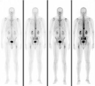
Figure 132 is the bone scan of a 73-year-old man who is referred from his family doctor with diffuse bone pain, fatigue, and right knee pain. Examination is notable for pain with motion about the right knee and mild hyporeflexia.
..Further imaging shows pulmonary metastases without an obvious primary tumor of origin and an incomplete fracture of the right distal femur. A decision is made to surgically treat his distal femur fracture. What is the role of establishing a preoperative histologic diagnosis for this patient?
1) The distal femoral lesion should undergo needle biopsy first.
2) Bone marrow biopsy should be done first.
3) Preoperative biopsy is not necessary because a metastatic process is present.
4) Biopsy is not necessary if the lesion demonstrates a standardized uptake value (SUV) greater than 3 on fluorodeoxyglucose positron emission tomography (PET) imaging.
.
- CT scan of the chest, abdomen, and pelvis and laboratory studies. PREFERRED RESPONSE: 3- intravenous bisphosphonate treatment.
PREFERRED RESPONSE: 3- Preoperative biopsy is not necessary because a metastatic process is present
