العربية

Understanding Multiple Epiphyseal Dysplasia: Orthopedic Cases

Updated: Feb 2026 83 Views
Understanding Multiple Epiphyseal Dysplasia: Orthopedic Cases
📖 Clinical Article

ORTHOPEDIC CASES /MULTIPLE EPIPHYSEAL DYSPLASIA ## Skeletal Dysplasias with Predominantly Epiphyseal Involvement
1. ### Multiple Epiphyseal Dysplasia
Multiple epiphyseal dysplasia (MED) is characterized by the disturbance of enchondral ossification involving numerous epiphyses. MED is usually transmitted in an autosomal dominant manner, although autosomal recessive transmission has also been reported. Different levels of deformities may be present in one patient. Usually lower extremity joint pain with decreased range of motions and limping are the main complaints. Dominantly hips, knees, and ankles are affected. Irregular, fragmented epiphyses and flat articular surfaces with normal metaphyses and mild shortening of the tubular bones can be observed.
Upper extremity involvement may differ from minimal to severe with significant deformities (Figs. 1.1–1.8).

Illustration 1 for Understanding Multiple Epiphyseal Dysplasia: Orthopedic Cases
🔍 Click to enlarge
Clinical Radiograph / Orthopedic Image

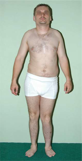
--- Fig. 1.1 Normal or moderately short height with normal proportions
Illustration 2 for Understanding Multiple Epiphyseal Dysplasia: Orthopedic Cases
🔍 Click to enlarge
Clinical Radiograph / Orthopedic Image

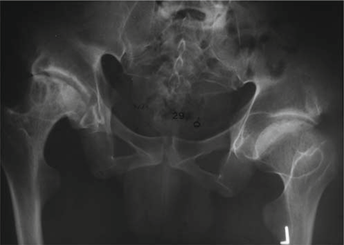
Fig. 1.2 Severely affected right hip with fragmentation of the epiphysis andflattening joint surfaces 2.
Illustration 3 for Understanding Multiple Epiphyseal Dysplasia: Orthopedic Cases
🔍 Click to enlarge
Clinical Radiograph / Orthopedic Image
Fig. 1.3 Normally developed knee joint with fragmentation and moderate deformity of the patella
Illustration 4 for Understanding Multiple Epiphyseal Dysplasia: Orthopedic Cases
🔍 Click to enlarge
Clinical Radiograph / Orthopedic Image
Fig. 1.4 Fingers are equally shortened
Illustration 5 for Understanding Multiple Epiphyseal Dysplasia: Orthopedic Cases
🔍 Click to enlarge
Clinical Radiograph / Orthopedic Image

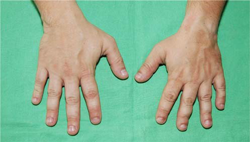
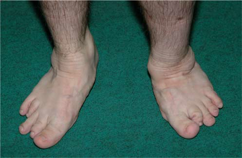
Fig. 1.5 Toes are variably shortened
Illustration 6 for Understanding Multiple Epiphyseal Dysplasia: Orthopedic Cases
🔍 Click to enlarge
Clinical Radiograph / Orthopedic Image
Fig. 1.6 Irregular proximal humeral epiphysis with large, flat articular surface
Illustration 7 for Understanding Multiple Epiphyseal Dysplasia: Orthopedic Cases
🔍 Click to enlarge
Clinical Radiograph / Orthopedic Image
Fig. 1.7 Bilateral irregular distal humeral epiphyses with deformity of the trochlea
Illustration 8 for Understanding Multiple Epiphyseal Dysplasia: Orthopedic Cases
🔍 Click to enlarge
Clinical Radiograph / Orthopedic Image

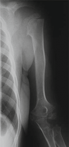
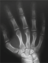
--- Fig. 1.8 The short tubular bones
of the hand are shortened without any significant deformity

Dr. Mohammed Hutaif
Written & Medically Reviewed by
Consultant Orthopedic & Spine Surgeon