Comprehensive Introduction and Patho-Epidemiology
Metacarpal fractures represent a profoundly significant proportion of skeletal trauma to the upper extremity, accounting for approximately 18% to 44% of all hand fractures encountered in acute orthopedic and plastic surgery practices. While the historical precedent for managing many isolated, closed metacarpal fractures has heavily favored non-operative modalities—such as cast immobilization or closed reduction and percutaneous pinning (CRPP)—the modern evolution of high-energy trauma mechanisms necessitates a more aggressive, surgically precise approach. High-velocity crush injuries, industrial accidents, and complex ballistic trauma frequently result in highly unstable, comminuted, or multi-segmental fracture patterns that demand rigid internal fixation. Open reduction and internal fixation (ORIF) utilizing modern plating systems provides unparalleled biomechanical stability, allowing for the meticulous anatomical restoration of digital length, axial alignment, and rotational congruity.
The overarching philosophy of metacarpal plate osteosynthesis extends far beyond mere bony union; it is fundamentally predicated on the facilitation of immediate, early active mobilization. The hand is an exquisitely unforgiving anatomical unit where prolonged immobilization invariably precipitates a cascade of debilitating complications, most notably extensor tendon adhesions, capsular contractures, and profound joint stiffness. By achieving absolute stability through rigid plate fixation, the orthopedic surgeon effectively bypasses the need for prolonged external splinting, thereby allowing the gliding planes of the intricate flexor and extensor mechanisms to be maintained during the critical early phases of bone healing.
However, the decision to proceed with open plate fixation is not benign and must be meticulously weighed against the inherent iatrogenic risks associated with the procedure. Extensive soft tissue dissection, aggressive periosteal stripping, and the introduction of prominent metallic hardware into the inherently thin dorsal soft tissue envelope of the hand can paradoxically induce the very stiffness the surgeon seeks to avoid. Mastery of metacarpal plating, therefore, requires a profound, almost intuitive understanding of hand biomechanics, precise and judicious implant selection, and flawless, tissue-respecting surgical execution.
The epidemiology of these fractures reveals a bimodal distribution, predominantly affecting young, active males in their second and third decades due to altercations, sports injuries, and occupational hazards, alongside an older demographic suffering from low-energy osteoporotic falls. The economic impact of these injuries is staggering, given the potential for prolonged absence from the workforce. Consequently, the primary objective of the operative surgeon is not merely radiographic union, but the rapid, complete restoration of the patient's functional capacity and pre-injury occupational status.
Detailed Surgical Anatomy and Biomechanics
Osteology and the Metacarpal Arch
The metacarpals form the foundational architectural framework of the hand, consisting of a proximal base articulating with the carpus, a diaphyseal shaft, a metaphyseal neck, and a distal articular head. The human hand is not a flat structural entity; it is organized into a complex, dynamic transverse and longitudinal arch system. The second and third metacarpals form the rigid central pillar of the hand, securely anchored to the trapezoid and capitate via robust ligamentous complexes, rendering them highly immobile. Conversely, the fourth and fifth metacarpals are highly mobile, articulating with the hamate to allow for the dynamic cupping of the palm necessary for a powerful grip. This anatomical dichotomy dictates surgical decision-making: the rigid central metacarpals tolerate very little angulation or shortening, whereas the mobile ulnar border metacarpals possess a greater capacity to compensate for minor residual deformities.
Musculotendinous Deforming Forces
Understanding the intrinsic and extrinsic musculotendinous forces acting upon the metacarpals is paramount for anticipating fracture displacement and achieving successful reduction. The intrinsic muscles, specifically the interossei and lumbricals, exert a profound deforming force on metacarpal shaft and neck fractures. Because these muscles originate on the metacarpal shafts and insert onto the proximal phalanges and extensor expansions, their contraction pulls the distal fracture fragment proximally and volarly. Concurrently, the extrinsic flexor tendons exert a powerful longitudinal compressive force that exacerbates shortening and volar angulation. This predictable interplay of forces almost universally results in an apex-dorsal angular deformity. If left uncorrected, this angulation alters the biomechanical mechanics of the extrinsic extensors, leading to a pseudo-claw deformity, extensor lag, and a functionally compromised grip.
The Tension Band Principle in Metacarpal Plating
The metacarpal shaft is subjected to highly complex, multi-planar forces during normal hand function, primarily driven by the powerful flexor tendons which exert a strong bending moment. This bending moment creates immense tensile forces along the dorsal cortex and corresponding compressive forces along the palmar (volar) cortex. To counteract these physiological forces, a plate applied to the dorsal surface functions fundamentally as a tension band. For this biomechanical principle to succeed, the palmar cortical buttress must be anatomically restored and structurally sound.
When the palmar cortex is intact or anatomically reduced, the dorsal plate is subjected primarily to tensile stress, while the bone itself absorbs the physiological compressive loads. This load-sharing construct is highly stable and conducive to primary bone healing. However, if the anterior buttress is deficient—due to severe volar comminution or an inadequate surgical reduction—the plate is forced to function as a load-bearing implant. Under these circumstances, the plate is subjected to cyclical bending stresses with every finger flexion, which will inevitably lead to implant fatigue, hardware failure, and nonunion.
Vascular Anatomy and Periosteal Preservation
The vascular supply to the metacarpals is derived from a dual system: the nutrient arteries entering the volar aspect of the diaphysis, and an extensive periosteal network supplied by the dorsal and palmar metacarpal arteries. In the context of trauma, the intramedullary blood supply is often disrupted by the fracture itself, rendering the bone highly dependent on the surrounding periosteal vascular network for survival and subsequent callus formation. Aggressive surgical stripping of the dorsal periosteum during plate application devascularizes the underlying cortex, significantly increasing the risk of delayed union, nonunion, and infection. Therefore, surgical exposure must be meticulously restricted to the absolute minimum required for anatomical reduction and implant placement.
Exhaustive Indications and Contraindications
The indications for plate fixation of the metacarpals are strictly defined to optimize functional outcomes in scenarios where less invasive methods—such as cast immobilization or percutaneous Kirschner wire fixation—would unequivocally fail to provide adequate stability. The decision matrix must incorporate fracture geometry, patient demographics, soft tissue integrity, and the anticipated compliance of the patient with rigorous postoperative rehabilitation protocols.
Primary indications include multiple metacarpal fractures, where the loss of the adjacent intact metacarpal's "splinting effect" necessitates rigid internal fixation to restore the architectural arch of the hand. Displaced diaphyseal fractures, specifically transverse, short oblique, or short spiral configurations that cannot be reduced or maintained by closed means, are classic indications. Furthermore, intraarticular and periarticular fractures involving the metacarpophalangeal (MCP) or carpometacarpal (CMC) joints require precise anatomical reconstruction of the articular surface to prevent post-traumatic osteoarthritis, a goal best achieved through direct visualization and rigid micro-fixation.
Conversely, absolute and relative contraindications must be rigorously respected. Severe soft tissue compromise, such as massive degloving injuries or active deep space infections, precludes the immediate use of internal hardware due to the unacceptably high risk of catastrophic deep infection and subsequent osteomyelitis. In such devastating scenarios, external fixation or delayed definitive management is mandatory. Extreme comminution where bridging is impossible without massive structural bone grafting may also warrant alternative stabilization strategies.
| Clinical Parameter | Indications for Plate Fixation | Contraindications for Plate Fixation | Rationale / Clinical Context |
|---|---|---|---|
| Fracture Pattern | Multiple adjacent metacarpal fractures; displaced transverse/short oblique; intra-articular step-off >1mm. | Undisplaced fractures; stable extra-articular fractures reducible by closed means. | Plating restores the structural arch when the "splinting effect" of adjacent bones is lost. Stable fractures do not warrant the soft tissue trauma of ORIF. |
| Deformity / Alignment | Shortening >2-5mm; Malrotation of any degree; Apex dorsal angulation >10° (index/long) or >30° (ring/small). | Acceptable angulation in border digits without clinical malrotation or functional deficit. | Rotational deformity is poorly tolerated; 5° of malrotation causes significant digital overlap. Mobile ulnar digits tolerate more angulation than rigid radial digits. |
| Soft Tissue Envelope | Intact dorsal soft tissues; clean, sharply demarcated open wounds (Gustilo-Anderson Type I/II). | Severe crush injuries with compromised vascularity; massive degloving; active gross infection. | Plating requires a viable soft tissue envelope to cover the hardware and prevent devascularization/infection. External fixation is preferred in severe trauma. |
| Bone Quality | Good cortical density capable of achieving secure screw purchase (bicortical fixation). | Severe osteopenia/osteoporosis where screw pull-out is highly probable. | Poor bone stock may necessitate locking plates, intramedullary devices, or acceptance of minor deformity over hardware failure. |
| Patient Factors | High-demand patients (manual laborers, athletes); compliant with early active motion protocols. | Medically unstable patients; severe non-compliance; profound baseline dementia. | The primary benefit of plating is early motion. If the patient cannot participate in therapy, the risks of plating (adhesions/stiffness) outweigh the benefits. |
💡 Clinical Pearl: Assessing Malrotation
Rotational deformity is the absolute least tolerated malalignment in metacarpal fractures. A mere 5 degrees of malrotation at the metacarpal base can translate to 1.5 cm of digital overlap at the fingertips during flexion, severely compromising grip strength and fine motor mechanics. Always assess the digital cascade intraoperatively by passively flexing the wrist to induce the tenodesis effect; the digits should naturally point toward the scaphoid tubercle without overlapping.
Pre-Operative Planning, Templating, and Patient Positioning
Advanced Imaging and Pre-Operative Templating
Meticulous preoperative planning is the cornerstone of successful metacarpal osteosynthesis. High-quality, orthogonal radiographs—comprising posteroanterior (PA), true lateral, and semi-pronated oblique views—are mandatory. The true lateral view is particularly critical, albeit difficult to interpret due to the superimposition of adjacent metacarpals; it is essential for assessing the degree of apex-dorsal angulation and the integrity of the volar cortex. For complex intra-articular fractures of the metacarpal head or base (such as Rolando variants), a fine-cut computed tomography (CT) scan with 3D reconstructions is highly recommended to precisely map the fracture lines, identify occult articular step-offs, and plan the trajectory of interfragmentary lag screws.
Digital templating should be performed to anticipate implant size, screw length, and plate configuration. The surgeon must evaluate the fracture geometry to determine whether the construct will rely on absolute stability (compression plating for transverse fractures, lag screw with neutralization plate for oblique fractures) or relative stability (bridge plating for highly comminuted segments). Having a comprehensive array of mini-fragment systems—typically 1.5-mm, 2.0-mm, and 2.4/2.7-mm sets—available in the operating theater is non-negotiable, as intraoperative findings frequently necessitate a deviation from the primary preoperative plan.
Implant Selection Considerations
The evolution of low-profile, anatomically contoured titanium and stainless-steel plating systems has revolutionized hand trauma surgery.
* 2.4-mm and 2.7-mm Dynamic Compression Plates (DCP): Utilized for robust fixation across transverse diaphyseal fractures in larger patients where maximum rigidity is required.
* 1/4 and 1/3 Tubular Plates: These are less bulky and ideal for stable fractures or as strut plates in cases of segmental bone loss. Their eccentric screw holes allow for dynamic compression, though they lack the inherent strength of a true DCP.
* Locking Plate Technology: Fixed-angle locking plates are increasingly utilized, particularly in osteoporotic bone or severely comminuted metaphyseal fractures where traditional screw purchase is compromised. The locking head screws thread directly into the plate, creating a rigid single-beam construct that does not rely on friction between the plate and the bone, thereby preserving periosteal blood supply.
* Specialty Plates (T-Plates, L-Plates, Condylar Plates): Specifically designed for periarticular and metaphyseal fractures where multiple points of fixation are required in a very short bone segment, particularly around the MCP and CMC joints.
Patient Positioning and Anesthesia
The patient is positioned supine on the operating table with the operative arm extended onto a radiolucent hand table. The height of the table must be adjusted to ensure ergonomic comfort for the operating surgeon and the assistant, who will be seated opposite one another. A well-padded pneumatic tourniquet is applied to the proximal arm.
Anesthesia typically consists of a regional brachial plexus block (axillary or supraclavicular) combined with intravenous sedation, which provides excellent intraoperative muscle relaxation and prolonged postoperative analgesia. Prophylactic intravenous antibiotics (typically a first-generation cephalosporin) are administered at least 30 minutes prior to tourniquet inflation. The arm is exsanguinated utilizing an Esmarch bandage, and the tourniquet is inflated to 250 mmHg or 100 mmHg above the patient's systolic blood pressure. Tourniquet time must be meticulously monitored, with a hard limit of 120 minutes before a deflation period is mandated.
Step-by-Step Surgical Approach and Fixation Technique
The Dorsal Surgical Approach
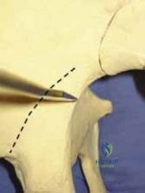
- Incision Planning: A longitudinal or slightly curvilinear dorsal incision is utilized. For isolated metacarpal fractures, the incision is centered directly over the palpable dorsal ridge of the affected bone. When addressing adjacent multiple metacarpal fractures (e.g., the 3rd and 4th metacarpals), a single longitudinal incision placed precisely in the intermetacarpal space is strongly advocated. This allows for the mobilization of broad fasciocutaneous flaps to access both bones, significantly minimizing cumulative soft tissue trauma and reducing the risk of ischemic skin bridge necrosis.
- Superficial Dissection: The subcutaneous tissues are sharply and bluntly dissected. Extreme vigilance must be maintained to identify, meticulously dissect, and retract the dorsal sensory branches of the radial nerve (radially) and the ulnar nerve (ulnarly). Concurrently, the dorsal venous network must be preserved whenever anatomically feasible to prevent postoperative venous congestion and massive digital edema, which severely impedes rehabilitation.
- Extensor Mechanism Management: The extensor digitorum communis (EDC) tendon is identified. Depending on the specific fracture location, the tendon can be retracted laterally (usually radially to exploit the natural laxity) or, less favorably, split longitudinally down its midline. Splitting the tendon provides direct access to the diaphysis but significantly increases the risk of postoperative tendon adhesions and should be reserved for cases where lateral retraction provides inadequate exposure.
- Periosteal Stripping: The dorsal periosteum is incised longitudinally. Using a sharp Freer elevator, the periosteum is elevated just enough to expose the fracture surfaces and accommodate the width of the chosen plate. Aggressive, circumferential periosteal stripping must be strictly forbidden, as it obliterates the vascular supply critical for osteogenesis.
⚠️ Surgical Warning: Soft Tissue Handling
Aggressive periosteal stripping and excessive, heavy-handed retraction of the extensor mechanism are the primary iatrogenic culprits for postoperative tendon adhesions, complex regional pain syndrome (CRPS), and profound joint stiffness. Expose only what is absolutely necessary for anatomical reduction and plate application. Respect the soft tissue envelope above all else.
1. Fracture Reduction and Provisional Fixation
Anatomical reduction is the absolute prerequisite for successful plating. The fracture site is meticulously cleared of hematoma, fibrin debris, and interposed soft tissue (often the interosseous muscle belly) using a dental pick and copious irrigation.
Achieving and maintaining provisional fixation is frequently the most challenging aspect of the procedure. Standard reduction forceps are often too bulky for the tight intermetacarpal spaces of the central digits. It is highly recommended to utilize 0.045-inch or 0.062-inch Kirschner wires placed percutaneously or directly into the fragments as "joysticks" to manipulate the bone. Once reduced, the fracture can be provisionally stabilized with a trans-fixation K-wire placed outside the planned footprint of the plate. Alternatively, an experienced assistant can manually hold the reduction and the pre-contoured plate firmly against the metacarpal dorsum using a Freer elevator while the primary surgeon performs the initial drilling.
2. Plating Transverse Diaphyseal Fractures (Compression Plating)
For transverse fractures where an adequate palmar cortical buttress has been anatomically restored:
1. Select an appropriately sized 2.0-mm or 2.4-mm dynamic compression plate.
2. Plate Contouring: Contour the plate to perfectly match, or slightly exceed, the natural dorsal bow of the metacarpal. Pre-bending the plate ensures that as the eccentric screws are tightened, the fracture site is compressed uniformly across both the dorsal and palmar cortices, preventing palmar gapping which leads to cyclical failure.
3. Apply the plate dorsally to act as a tension band.
4. Drilling and Tapping: Drill the first hole adjacent to the fracture line in a neutral position. Measure the depth, tap if using non-self-tapping screws, and insert the screw but do not fully tighten.
5. Dynamic Compression: Drill the corresponding hole on the opposite side of the fracture in an eccentric position (away from the fracture line). As this screw is driven home, the spherical head engages the ramped plate hole, dynamically shifting the bone fragment and compressing the fracture.
6. Screw Tightening Protocol: Tighten all screws terminally using the force of only two or three digits on the screwdriver handle. The diaphyseal cortex of the metacarpal is relatively thin; over-tightening with a full-hand power grip will easily strip the thread purchase, catastrophically compromising the construct. Ensure screw purchase in at least four cortices (two bicortical screws) distal and proximal to the fracture zone.
3. Plating Short Oblique and Spiral Fractures (Lag Screw and Neutralization)
Short oblique and spiral fractures are inherently unstable, highly prone to shortening, and notorious for inducing malrotation.
1. Interfragmentary Lag Screw: First, achieve perfect anatomical reduction. Drill a gliding hole in the near cortex and a thread hole in the far cortex. Place a 1.5-mm or 2.0-mm interfragmentary lag screw precisely perpendicular to the fracture plane. This provides absolute stability and powerful interfragmentary compression.
2. Neutralization Plate: The lag screw alone cannot withstand physiological loading. Apply a dorsal plate (e.g., a 1/4 tubular or 2.0-mm locking plate) spanning the fracture. This plate functions purely to neutralize rotational, bending, and shearing stresses that would otherwise cause the isolated lag screw to fail.
4. Plating Intraarticular and Periarticular Fractures
Metaphyseal and intraarticular fractures require specialized implants like T-shaped, oblique L-plates, or condylar plates.
1. Articular Reconstruction: For intraarticular fractures (e.g., metacarpal head), the joint capsule must be opened (arthrotomy) to directly visualize the articular surface. Anatomically reduce the articular fragments and lag them together using a countersunk mini-screw placed perpendicular to the fracture site.
2. Plate Application Sequence: When utilizing a T-plate or L-plate for metaphyseal fractures, always apply the transverse side arm(s) to the short metaphyseal fragment first. If the diaphyseal shaft is secured first, driving screws into the transverse arms can draw the underlying, often slightly offset metaphyseal bone fragment up to the plate asymmetrically, instantly inducing an iatrogenic rotational deformity.
🔪 Surgical Pitfall: Distal Metaphyseal Plating
With distal metaphyseal metacarpal fractures, standard dorsal plating frequently interferes with the gliding of the central slip of the extensor mechanism, leading to severe extension lag, chronic tenosynovitis, or outright tendon rupture.
Solution: Utilize a specialized 2.0-mm condylar plate. Apply it dorsoradially or dorsoulnarly, securing the blade or transverse screws directly through the dorsal tubercle—the anatomical origin of the collateral ligament. This strategic placement avoids the central extensor apparatus entirely.
5. Managing Bone Loss and Segmental Defects (Bridge Plating)
High-energy ballistic or crush injuries with significant cortical substance loss require bridge plating to maintain digital length, axial alignment, and rotation while bypassing the zone of injury.
1. Utilize a robust plate (e.g., a 2.4-mm locking plate or a 2.7-mm DCP acting as an internal fixator).
2. Secure the plate proximally and distally with at least three bicortical screws per segment, completely bypassing the zone of comminution. Do not attempt to place screws into the devitalized intermediate fragments.
3. Because this construct is subjected to massive bending loads without the support of a palmar buttress, it is highly susceptible to fatigue failure.
4. Bone Grafting: Cancellous autograft (typically harvested from the distal radius via a Kurbs approach, or the iliac crest for larger defects) must be packed into the osseous defect. This can be performed acutely if the soft tissue bed is pristine, or in a staged procedure (Masquelet technique) if the wound is contaminated.
Complications, Incidence Rates, and Salvage Management
Despite flawless preoperative planning and meticulous intraoperative technique, the surgical management of metacarpal fractures is fraught with potential complications. The delicate interplay between the osseous architecture and the overlying tendinous structures means that even minor technical errors can yield profound functional deficits.
| Complication | Estimated Incidence | Etiology / Pathophysiology | Prevention and Salvage Management |
|---|---|---|---|
| Extensor Tendon Adhesions | 15% - 30% | Most common complication. Caused by aggressive periosteal stripping, prominent hardware, or delayed postoperative mobilization. | Prevention: Low-profile implants, minimal soft tissue handling, immediate active therapy. Salvage: Aggressive hand therapy; surgical tenolysis and hardware removal after complete bony union (typically >6-9 months post-op). |
| Hardware Prominence / Pain | 10% - 25% | The dorsal soft tissue envelope of the hand is exceptionally thin. Standard plates often become palpable and symptomatic during heavy grip. | Prevention: Countersink screws, utilize modern low-profile titanium systems. Salvage: Elective hardware removal once solid clinical and radiographic union is achieved. |
| Malunion (Malrotation) | 2% - 8% | Failure to assess the digital cascade intraoperatively; asymmetrical plate tightening; inadequate reduction of spiral fractures. | Prevention: Intraoperative tenodesis test; visual alignment of fingernails. Salvage: Corrective rotational osteotomy at the metacarpal base or diaphysis, followed by rigid re-plating. |
| Nonunion / Hardware Failure | 1% - 5% | Failure to restore the palmar cortical buttress, leading to cyclical bending and fatigue failure of the plate. Infection. Devitalized bone. | Prevention: Anatomical volar reduction; bridge plating with acute bone grafting for defects. Salvage: Revision ORIF with robust plating, autologous bone grafting, and rigorous infection workup. |
| Deep Space Infection | 1% - 3% | Contaminated open fractures; compromised soft tissue envelope; prolonged operative times; hematoma formation. | Prevention: Thorough debridement of open wounds; meticulous hemostasis; prophylactic antibiotics. Salvage: Urgent surgical I&D. Retain stable hardware until union if possible; remove loose hardware and place antibiotic spacers if necessary. |
| Complex Regional Pain Syndrome (CRPS) | 1% - 4% | Aberrant neuroinflammatory response to trauma and surgery. Exacerbated by poor pain control and prolonged immobilization. | Prevention: Gentle tissue handling, avoidance of nerve traction, early mobilization, adequate analgesia. Salvage: Multidisciplinary approach: aggressive therapy, neuropathic medications (gabapentin), sympathetic nerve blocks. |
Phased Post-Operative Rehabilitation Protocols
The primary, overriding advantage of rigid plate fixation is the ability to initiate early, safe motion. A delayed or excessively conservative rehabilitation protocol completely negates the purpose of the surgery and virtually guarantees a poor functional outcome. Rehabilitation must be closely supervised by a certified hand therapist (CHT) and tailored to the rigidity of the fixation achieved intraoperatively.
Phase I: Immobilization and Edema Control (Days 1 to 5)
Immediately postoperatively, the hand is placed in a bulky, compressive soft dressing and supported by a volar plaster or fiberglass splint in the classic "intrinsic-plus" (Edinburgh) position:
* Wrist extended 20 to 30 degrees to optimize the mechanical advantage of the flexors.
* MCP joints flexed 70 to 90 degrees. This is absolutely critical to maintain the collateral ligaments at their maximum length, preventing devastating extension contractures.
* Interphalangeal (IP) joints fully extended to prevent volar plate contractures.
The patient is instructed on strict elevation above heart level and active range of motion of the shoulder, elbow, and uninvolved digits to mitigate edema.
Phase II: Early Active Motion (Days 5 to 14)
At the first postoperative visit (typically days 3-5), the bulky surgical dressing is removed. The wound is inspected, and a custom, removable thermoplastic splint is fabricated by the hand therapist, maintaining the intrinsic-plus position between exercise sessions.
* Active Range of Motion (AROM): Gentle, active flexion and extension of the digits are initiated. Early gliding of the extensor tendons directly over the dorsal plate is the most critical factor in preventing dense, restrictive adhesions.
* Place-and-Hold Exercises: If the patient struggles with active initiation, the therapist may passively place the digits into flexion and ask the patient to actively maintain the position.
* Passive range of motion (PROM) is generally avoided at this stage to prevent excessive stress on the healing surgical wound and the fracture construct.
Phase III: Intermediate Rehabilitation (Weeks 2 to 6)
Sutures are typically removed between 10 and 14 days, provided the incision is clean and dry.
* Scar Management: Once the wound is fully healed, aggressive scar massage and silicone gel sheeting are initiated to mobilize the cutaneous tissues away from the underlying extensor mechanism.
* Progressive Motion: PROM is carefully introduced. If joint stiffness or extrinsic tightness is noted, gentle dynamic or static-progressive splinting may be implemented during the day or at night.
* Tendon Gliding Exercises: Hook fist, straight fist, and full fist exercises are heavily emphasized to ensure differential gliding of the flexor digitorum superficialis (FDS) and profundus (FDP) tendons.
Phase IV: Strengthening and Return to Function (Weeks 6 to 12)
At 6 weeks postoperatively, a comprehensive clinical and radiographic evaluation is performed to assess for callus formation, obliteration of the fracture lines, and overall consolidation.
* Once clinical union (absence of pain at the fracture site with stress) and radiographic union are evident, progressive strengthening exercises are commenced using putty, hand grippers, and weighted activities.
* Return to heavy manual labor, industrial work, or contact sports is typically restricted until 10 to 12 weeks postoperatively to ensure the bone has remodeled sufficiently to withstand peak physiological loads without refracture.
Summary of Landmark Literature and Clinical Guidelines
The contemporary approach to metacarpal plating is heavily informed by decades of biomechanical research and clinical outcomes studies. The foundational principles of rigid internal fixation in the hand were largely pioneered by the AO Foundation (Arbeitsgemeinschaft für Osteosynthesefragen), which established the absolute necessity of anatomical reduction, stable fixation, preservation of blood supply, and early active mobilization.
Landmark biomechanical studies by Hastings and others elucidated the profound impact of minor rotational deformities, establishing the rigid clinical threshold that even a few degrees of malrotation at the metacarpal base necessitates surgical correction. Furthermore, literature evaluating implant biomechanics has consistently demonstrated that dorsal plating provides superior resistance to apex-dorsal bending forces compared to intramedullary pinning or crossed K-wires, albeit at the cost of a higher complication profile regarding soft tissue adhesions.
Recent systematic reviews and meta-analyses comparing conventional non-locking plates to modern locking plate systems in the hand have shown that while locking plates provide superior biomechanical stability in osteoporotic bone or severely comminuted fracture models, they do not offer a statistically significant clinical advantage in terms of final range of motion or union rates in simple, transverse fractures in young, healthy patients. Furthermore, the increased profile and cost of locking systems must be judiciously considered. The consensus among leading academic hand surgeons remains that the meticulous handling of the soft tissue envelope and the rigorous adherence to early mobilization protocols are far more predictive of an excellent functional outcome than the specific brand or locking capability of the titanium implant utilized.
By adhering strictly to the biomechanical principles of tension band fixation, demonstrating profound respect for the delicate and unforgiving soft tissue envelope of the hand, and instituting early, expertly supervised rehabilitation, orthopedic surgeons can consistently achieve excellent, predictable functional outcomes in the management of complex metacarpal fractures.
This academic resource was prepared and medically reviewed by Prof. Dr. Mohammed Hutaif, Consultant Orthopedic & Spine Surgeon. It is formulated specifically for medical students, orthopedic residents, and surgeons preparing for high-stakes board examinations (AAOS, FRCS Tr & Orth, Arab Board).
