DEFINITION
This injury was initially reported by Giovanni Monteggia in 1814 as a fracture of the ulna associated with an anterior dislocation of the radial head. 6 The term “Monteggia lesions” was coined by Bado to describe any fracture of the ulna associated with a dislocation of the radiocapitellar joint. 1 The Bado classification of Monteggia lesions, 1 with the Jupiter subclassification of type II fractures,4 is shown in Table 1. Equivalent injuries in adults Variable pathology that is thought to be equivalent to injuries classified by the Bado system Equivalent injuries do not always fall within the traditional definition of a Monteggia fracture in that they do not always have a concomitant radiocapitellar dislocation. Therefore, it can be argued that these injuries are not necessarily equivalent to Monteggia fractures. Type I and II injuries are the only ones that have equivalent injury patterns. ## PATHOGENESIS The exact mechanism of injury for Monteggia fractures is controversial. Proposed mechanisms of injury for type I injuries include the following: Direct blow to the posterior aspect of the elbow Fall on outstretched arm with hyperpronated hand (forearm pronation levers radial head anteriorly) Fall on outstretched arm Violent contraction of biceps pulling radial head anteriorly Proposed mechanism for type II injuries: hypothesized to occur when a supination force tensions the ligaments that are stronger than bone Proposed mechanism for type III injuries: direct blow to the inside of the elbow with or without rotation ## PATIENT HISTORY AND PHYSICAL FINDINGS The initial examination should systematically evaluate: Skin integrity Neurovascular status of the extremity Bony injury Ulna fracture Injury pattern Noncomminuted Comminution Associated injury to key structural elements of the ulna (coronoid, olecranon) Radial head injury Isolated dislocation without fracture Radial head or neck fracture ## IMAGING AND OTHER DIAGNOSTIC STUDIES Plain radiographs ( FIG 1): Orthogonal radiographs of the elbow, forearm, and wrist are required. Ulna fracture is easily identified. Radial head fracture or dislocation can be subtle, especially if radial head dislocation reduces. Computed tomography (CT) scans can be helpful to determine the extent of the bony injury and the location of fracture fragments. They are particularly helpful in fractures involving the coronoid, olecranon, and radial head. 3D CT reconstructions provide information on the spatial relationship of fracture fragments in comminuted fractures. ## DIFFERENTIAL DIAGNOSIS Isolated ulna fracture Nightstick fracture Olecranon fracture Fracture-dislocation of the elbow (“terrible triad” injury) Transolecranon fracture-dislocation ## NONOPERATIVE MANAGEMENT Monteggia fracture-dislocations in the adult population are generally treated surgically. Improved fixation methods and surgical technique have remarkably improved the results of surgery, making it a more reliable treatment option. ## SURGICAL MANAGEMENT
Preoperative Planning The timing of surgery depends on the condition of the soft tissues and the availability of necessary equipment and personnel. The surgeon should define all injuries that need to be addressed. Equipment requirements: Small fragment plates and screws or anatomic plating system Minifragment system Threaded Kirchner wires Radial head replacement Bone graft (allograft or autograft) ## Patient Positioning Lateral decubitus position with the arm over a padded arm support ( FIG 2) Supine positioning is an alternative approach (although it is not preferred because of difficulty in maintaining the arm across the chest). If this approach is used, a saline bag under the ipsilateral shoulder will help keep the arm across the chest. 303 ## Table 1 Bado Classification of Monteggia Lesions, With Jupiter Subclassification of Type II Fractures
Type Description
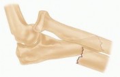
Illustration I Anterior dislocation of the radial head with fracture of the diaphysis of the ulna with anterior angulation of the ulna fracture (most common type of lesion) II Posterior or posterolateral dislocation of the radial head with fracture of the ulnar diaphysis with posterior angulation of the ulna fracture IIA Fracture at the level of the trochlear notch (ulna fracture involves the distal part of the olecranon and coronoid) IIB Ulna fracture is at the metaphyseal-diaphyseal junction, distal to the coronoid
 ---
---
 ---
---
 ---
---
 ---
---
 ---
---
 ---
---
 ---
---
 --- IIC Ulna fracture is diaphyseal IID Comminuted fractures involving more than one region 1. Lateral or anterolateral dislocation of the radial head with fracture of the ulnar metaphysis 2. Anterior dislocation of the radial head with a fracture of the proximal third of the radius and ulna at the same level Adapted from Bado J. The Monteggia lesion. Clin Orthop Relat Res 1967;50:717; and Jupiter JB, Leibovic SJ, Ribbans W, et al. The posterior Monteggia lesion. J Orthop Trauma 1991;5:395-402. 304
 ---
---
 ### FIG 2 • Lateral decubitus positioning is preferred.
 ---
---
 ### FIG 1 • Plain AP and lateral radiographs typically demonstrate fracture pattern. 305 ## TECHNIQUES
-
Surgical Approach A midline posterior skin incision is placed lateral to the tip of the olecranon ( TECH FIG 1A). Subcutaneous flaps are elevated on the fascia of the forearm. The medial antebrachial cutaneous nerve does not need to be identified if dissection is performed on the fascia of the flexor-pronator muscles since it is mobilized with the medial skin fla The interval between the flexor carpi ulnaris (FCU) and anconeus is developed along the subcutaneous border of the ulna to expose the fracture site. The amount of dissection required for exposure is dictated by the fracture pattern and the type of fixation to be used ( TECH FIG 1B).
 ---
---
 ### TECH FIG 1 • A. Posterior midline incision positioned just off the lateral aspect of the olecranon. B. Deep surgical interval uses the internervous plane between the anconeus and flexor carpi ulnaris. C. Exposure of the radial head can be accomplished by releasing the anconeus from the humerus and reflecting it proximally to expose the radial head. If the radial head needs to be addressed surgically, the anconeus can be mobilized more extensively through a Boyd approach ( TECH FIG 1C). If the ulna fracture permits, the radial head can be fixed through the fracture bed of the ulna before definitive fixation of the ulna. Once the ulna is fixed, access to the radial head is not possible. 1. ##
Radial Head Management
Radial head fractures are typically fixed before the ulna fracture is addressed. If the lesser sigmoid notch of the ulna is involved, determining radial length if radial head replacement is required can be difficult. Therefore, fractures are generally fixed before the ulna while replacement may need to be performed after ulnar fixation is completed in order to establish appropriate radial head sizing. Reconstructable fractures of the radial head are fixed ( TECH FIG 2A,B). Unreconstructable fractures of the radial head are replaced (TECH FIG 2C).
### TECH FIG 1 • A. Posterior midline incision positioned just off the lateral aspect of the olecranon. B. Deep surgical interval uses the internervous plane between the anconeus and flexor carpi ulnaris. C. Exposure of the radial head can be accomplished by releasing the anconeus from the humerus and reflecting it proximally to expose the radial head. If the radial head needs to be addressed surgically, the anconeus can be mobilized more extensively through a Boyd approach ( TECH FIG 1C). If the ulna fracture permits, the radial head can be fixed through the fracture bed of the ulna before definitive fixation of the ulna. Once the ulna is fixed, access to the radial head is not possible. 1. ##
Radial Head Management
Radial head fractures are typically fixed before the ulna fracture is addressed. If the lesser sigmoid notch of the ulna is involved, determining radial length if radial head replacement is required can be difficult. Therefore, fractures are generally fixed before the ulna while replacement may need to be performed after ulnar fixation is completed in order to establish appropriate radial head sizing. Reconstructable fractures of the radial head are fixed ( TECH FIG 2A,B). Unreconstructable fractures of the radial head are replaced (TECH FIG 2C).
 ---
---
TECH FIG 2 • A,B. Preoperative and postoperative radiographs demonstrating open reduction and internal fixation of the radial head component of the Monteggia fracture.
(continued)
306
 ---
---
TECH FIG 2 • (continued) C. Postoperative radiograph of a Monteggia fracture in which the radial head fracture needed to be replaced.
-
** Ulna Fracture Fixation ### No Articular Involvement of the Ulnohumeral Joint Ulna fractures distal to the coronoid can be plated laterally or on the subcutaneous border of the ulna. Lateral plate placement is preferred by some to prevent hardware prominence. ### Articular Involvement of the Ulnohumeral Joint Fractures extending proximal to the coronoid require the plate be placed on the subcutaneous border of the ulna to accommodate the complex geometry of this region. In general, the ulna fracture is reconstructed from distal to proximal. Ensure that any associated injury to the coronoid is identified and addressed. The fracture is reconstructed by fixing the distal fragments; this may require interfragmentary fixation or subarticular Kirchner wires ( TECH FIG 3A). As fixation progresses proximally, reconstruction of the coronoid and greater sigmoid notch is performed. Particular attention is directed at anatomic reconstruction of the articular surface. Coronoid involvement with a Monteggia fracture-dislocation often extends distally into the volar cortex of the ulna, as opposed to the axial-plane fracture patterns characterized by Regan and Morrey 9 ( TECH FIG 3B). Larger fragments can be definitively fixed with antegrade lag screws from the dorsal aspect of the ulnar or can be provisionally fixed with threaded wires and ultimately definitively fixed once the plate is applied to the dorsal aspect of the ulna. Coronoid fracture exposure can typically be obtained through the olecranon fracture. If this does not provide sufficient exposure, the FCU can be elevated from the dorsal aspect of the ulna. The final fragment to be fixed is the olecranon fragment. The attached triceps will obscure fracture reduction if reduced before distal reconstruction ( TECH FIG 3C). Definitive fixation is performed with a dorsal plate. The triceps is partially split to allow the proximal aspect of the plate to oppose the olecranon ( TECH FIG 3D).
 ---
---
 ### TECH FIG 3 • A. Monteggia fractures with articular involvement should be fixed distal to proximal. Fixation may require intramedullary Kirschner wires or interfragmentary fixation. B. Coronoid fracture often extends into the volar cortex of the ulnar.
(continued)
307
### TECH FIG 3 • A. Monteggia fractures with articular involvement should be fixed distal to proximal. Fixation may require intramedullary Kirschner wires or interfragmentary fixation. B. Coronoid fracture often extends into the volar cortex of the ulnar.
(continued)
307
 ** ---
TECH FIG 3 • (continued) C. The olecranon fragment with attached triceps is reduced and provisionally held with medial and lateral Kirschner wires pending definitive fixation. D. Final fixation for most Monteggia fractures is with a rigid plate applied to the dorsal cortex.
- ** Wound Closure The tourniquet is deflated and hemostasis is obtained. The fascia between the FCU and anconeus is closed with interrupted absorbable 0 or 1 suture. Subcutaneous tissues are closed with 3-0 absorbable suture and skin is closed with staples. I prefer to close the wound over a drain placed in the subcutaneous tissues to avoid hematoma. A well-padded dressing is applied and an anterior splint is placed with the elbow in full extension. PEARLS AND PITFALLS **--- | Indications ▪ Monteggia fracture-dislocations in adults require surgical intervention. Goals of ▪ The first goal is to restore ulnar length and location of the radial head. When the treatment articulation is involved, the goal is to obtain a concentric reduction with sufficient elbow stability that early range of motion is possible. 1. The second goal is to avoid complications that compromise function. Ulna ▪ Fractures distal to the coronoid need only to be fixed such that ulnar length is re-fractures established. 1. When plating these fractures, avoiding malreduction of the ulna is critical to reduction of the radial head. Failure to re-establish ulnar geometry can result in persistent subluxation or dislocation of the radial head ( FIG 3). 2. Fractures involving the articulation require stable fixation to re-establish a competent joint. Radial ▪ Radial head fractures are fixed or replaced. head Physical ▪ Early range of motion is the goal of treatment but may be delayed if fixation is therapy questionable. | --- | FIG 3 • Malunion of the ulna with resulting apex dorsal angulation results in dislocation of the radial head. | ---|---|--- 308 ## POSTOPERATIVE CARE The arm is splinted in full extension to take pressure off the posterior soft tissues. If a drain is used, the splint and dressing are removed when the drain output is less than 30 mL in 8 hours. If no drain is used, the dressing is removed on postoperative day 1. Active or active-assisted flexion and gravity-assisted extension is begun once the surgical dressings are removed. If fixation is tenuous because of poor-quality bone or comminution, mobilization is delayed. ## OUTCOMES Historically, the results of operative treatment of Monteggia fracture-dislocations have been unpredictable. 3, 7, 8, 11 The advent of rigid internal fixation has improved the results of operative treatment. 2, 4, 7 Certain factors have been associated with a poor clinical result 5: Bado type II injury Jupiter type IIa injury Fracture of the radial head Coronoid fracture Complications requiring further surgery ## COMPLICATIONS Complications associated with Monteggia fracturedislocations occur with frequency. A multicenter study evaluating Monteggia fracture-dislocations in adults demonstrated complications in 43% of the patients treated, with an unsatisfactory outcome in 46% of the patients treated. 10 Radial nerve palsy Most commonly posterior interosseous nerve Causes of injury include: Compression at the arcade of Frosche Direct trauma Traction with lateral displacement of the radial head Most common with type III fractures Complete resolution typically occurs. Malunion Most common in type II fractures with volar comminution that is not appreciated or addressed If radial head subluxation persists, malunion must be considered. Nonunion Causes of nonunion include: Infection Inadequate internal fixation Compression plate fixation required, particularly if fracture is comminuted Semitubular and reconstruction plates are not structurally strong enough. Radioulnar synostosis Seen with high-energy injuries with associated comminution Higher incidence if radial head fracture associated with ulna fracture at the same level Boyd approach implicated since the radius and ulna are exposed through the same incision ## REFERENCES 1. Bado J. The Monteggia lesion. Clin Orthop Relat Res 1967;50:71. 2. Boyd H, Boals J. The Monteggia lesion: a review of 159 cases. Clin Orthop Relat Res 1969;66:94-100. 3. Bruce H, Harvey JJ, Wilson JJ. Monteggia fractures. J Bone Joint Surg Am 1974;56A:1563-1576. 4. Jupiter JB, Leibovic SJ, Ribbans W, et al. The posterior Monteggia lesion. J Orthop Trauma 1991;5:395-402. 5. Konrad GG, Kundel K, Kreuz PC, et al. Monteggia fractures in adults: long-term results and prognostic factors. J Bone Joint Surg Br 2007;89B:354-360. 6. Monteggia GB. Instituzioni Chirurgiche. 2nd ed. Milan: G. Masperp, 1813-1815. 7. Reckling F. Unstable fracture-dislocations of the forearm (Monteggia and Galeazzi lesions). J Bone Joint Surg Am 1982;64A:857-863. 8. Reckling FW, Cordell LD. Unstable fracture-dislocations of the forearm: the Monteggia and Galeazzi lesions. Arch Surg 1968;96:999-1007. 9. Regan W, Morrey B. Fractures of the coronoid process of the ulna. J Bone Joint Surg Am 1989;71A:1348-1354. 10. Reynders P, De Groote W, Rondia J, et al. Monteggia lesions in adults: a multicenter Bota study. Acta Orthop Belg 1996;62(Suppl 1):78-83.
- Speed J, Boyd H. Treatment of fractures of ulna with dislocation of the head of radius (Monteggia fracture). JAMA 1940;115:1699-1705.
