Chapter
25
## Meniscoplasty for Discoid Lateral Meniscus
Jay C. Albright
DEFINITION
1. A discoid meniscus is abnormal in both thickness and amount of covering or interposition of the compartment or plateau.
2. Over 99% of cases occur on the lateral side of the knee, with an overall incidence of 1% to 15% of the general population.
3. Ten percent of children found to have a discoid meniscus will have it bilaterally.
ANATOMY
4. Three types of discoid meniscus are described: complete (covering entire compartment), incomplete (on partial compartment covering), and Wrisberg (complete or incomplete compartment covering with no peripheral attachments.5
5. Wrisberg type is by definition unstable, allowing displacement, popping, and locking as well.
PATHOGENESIS
6. It arises either congenitally or through abnormal development. No cases have been found in autopsies of fetal deaths or stillborns.
NATURAL HISTORY
7. Discoid menisci have frequently been found at autopsy in elderly, reportedly asymptomatic people.
8. Frequently it is an incidental finding.
9. Symptoms typically present in the late first or early second decade of life but may occur at any age.6
10. Symptoms are pain with or without loss of motion.
PATIENT HISTORY AND PHYSICAL FINDINGS
11. The common presentation is a young child (younger than 10 years) with a catch or popping of the lateral side of the knee with motion, with or without pain.
12. Some patients describe true mechanical locking symptoms.
13. The patient may present with painful or painless loss of motion.
14. The clinical examination may show a hypermobile lateral meniscus with palpable, audible, and frequently visual meniscal instability.
15. Effusion is a common finding. Objective signs of swelling with or without activity indicate irritation of the joint and possible tearing.
16. Loss of extension and joint line tenderness are also common.4
17. A discoid meniscus with a tear or instability will click or pop and may be uncomfortable. The results of the McMurray test will help with diagnosis.
1. Positive: pain and a pop or click
2. Negative: no pain and no pop or click
3. Equivocal: pop or click or pain without the other
18. Significant mobility of the lateral meniscus, while not uncommon, normally may indicate a discoid meniscus.
1. In children, varus instability may be due to accommodation of the large discoid lateral meniscus. Collateral ligament test results are important.
1. Normal: symmetric to the opposite side
2. Mild: 1 to 3 mm of increased laxity from the opposite side
IMAGING AND OTHER DIAGNOSTIC STUDIES
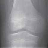
2. Radiographs may show flattening or sloping of the lateral femoral condyle, with widening of the lateral compartment compared to the medial compartment (
FIG 1A
).
3. MRI will show the discoid meniscus the best (
FIG 1B
).
1. A discoid meniscus will be thicker and wider than a normal meniscus.
2. Frequently signal change is present in the center of the discoid meniscus; this could represent a tear or degenerative tissue.1
3. There should be no more than three consecutive 3-mm cuts of the body of a meniscus on the sagittal view before it is separated into an anterior and posterior horn. Coronal cuts may also show a wide, thickened meniscus (more than 12 to 15 mm).
A

B

FIG 1 • A. Radiographs may show no significant changes, although there may be a widened lateral joint on weight-bearing views, and relative flattening of the lateral femoral condyle may be present. B. MRI shows the discoid meniscus clearly with a thickened, wide meniscus that also has abnormal signal intensity throughout the lateral meniscus. 1183

---
1184
Part 4
PEDIATRICS •
Section II
ARTHROSCOPIC AND SPORTS MEDICINEDIFFERENTIAL DIAGNOSIS
1. Meniscal cyst
2. Tear in a normal meniscus
3. Anterior cruciate ligament tear
4. Hypermobile lateral meniscus
5. Osteochondritis dissecans
6. Patellofemoral instability or dislocation
NONOPERATIVE MANAGEMENT
7. If there is no loss of motion or locking, a period of nonoperative management is the first line of defense.
8. Nonoperative treatment consists of activity modifications, anti-inflammatory medications, and swelling control (ice, elevation, and compression).
9. Patients with intermittent symptoms only that can be controlled with mild doses of nonsteroidal anti-inflammatories are candidates for nonoperative management.
SURGICAL MANAGEMENT
10. If locking, loss of motion, or persistent pain and disability exists despite nonoperative management, surgical intervention is indicated.3
Preoperative Planning
11. The surgeon should review imaging studies to evaluate the likelihood of a tear or the presence of other pathology.
12. The knee examination is repeated under anesthesia, including ligamentous testing, range of motion, and the McMurray test to evaluate whether significant lateral meniscal instability is present.
13. May indicate higher likelihood of the Wrisberg type of discoid meniscus.
Positioning
14. The patient is positioned supine.
15. A tourniquet is placed on the proximal thigh of the operative leg over padding.
16. A leg holder is placed over the tourniquet.
17. The opposite leg is padded and is placed in slight flexion at the hip.
18. The foot of the bed is flexed 90 degrees, allowing both legs to flex 90 degrees over the edge of the table.
Approach
19. Three standard arthroscopic portals are established with a no. 11 blade through stab incisions: inferolateral parapatellar portal for scope visualization, inferomedial parapatellar portal for instruments, and lateral suprapatellar pouch portal for outflow.
20. An accessory anterolateral portal may be established for another working portal.
21. If the remnant of the discoid meniscus is unstable or torn, requiring fixation or stabilization, a posterolateral approach should be made for inside-out suture fixation.
22. A lateral incision is made from the joint line distally by 2 cm, longitudinally in line with the posterior aspect of the fibu-lar head.
23. The interval between the biceps femoris and the iliotibial band is entered, as is the space deep to the lateral head of the gastrocnemius.
24. A posterior knee retractor is placed in this interval as far me-dially as possible to protect the neurovascular bundle.


TECHNIQUES
ARTHROSCOPIC SAUCERIZATION OF A DISCOID LATERAL MENISCUS
- After systematic arthroscopic evaluation of the knee is performed, the lateral compartment is opened in the figure 4 position.
- The type of discoid meniscus is determined using a probe sequentially over and under the posterior horn of the meniscus, pulling forward to evaluate displacement.
- Displacement of more than 40% to 50% anteriorly is unstable and requires stabilization with suture fixation.
- Determining peripheral instability may be difficult until the meniscoplasty is at least partly completed.
- Starting in the notch, the free edge of the discoid meniscus is identified ( TECH FIG 1A–C ).
- At this point an arthroscopic basket or a meniscal knife can be used to incise and remove the meniscus coronally from the notch toward the body of the meniscus.
- The surgeon should stop about 15 mm from the lateral edge of the meniscus to leave ample residual rim.
- A combination of arthroscopic baskets (angled, straight, up-biters, back-biters, and 90-degree side-biters) and shaver is employed to piecemeal the posterior and anterior aspects of the discoid meniscus ( TECH FIG 1D–G ).
- A meniscal rim of about 15 mm is maintained.
-
Attempts to thin the remainder of the thickened remnant should be done with care but can be performed with an aggressive shaver, baskets, or both.2
A B CTECH FIG 1•
Complete discoid lateral meniscus, visualization, and probing of anterior cruciate ligament (
A
); complete discoid with a tear visualized through the notch (
B
).
C.
Evaluation of the depth of the tear.
(continued)


Chapter 25 MENISCOPLASTY FOR DISCOID LATERAL MENISCUS1185D | E |
F
--- TECHNIQUESG | TECH FIG 1 • (continued) D. Initiation of saucerization through access point of the notch. E. Use of a shaver to remove loose pieces as well as shape the meniscus. F. Final appearance after saucerization. G. When the meniscus is unstable, suture techniques may be necessary for stabilization, as demonstrated with repeat probing after one all-inside device was needed to stabilize this meniscus.ALTERNATIVE TECHNIQUE FOR MENISCOPLASTY visualization to ensure that there is no inadvertent dam- edge of the discoid meniscus closer to the leading edge
age of the peripheral meniscus. of the incised meniscus.
arthroscopic locking grasper through the medial portal. meniscus.
sory lateral portal, ideally with a protective cannula or discoid, leaving the posterior portion of the discoid left
a sheath. to finish.
terior notch, leaving about 15 mm of anterior rim, di- rior aspect of the discoid with arthroscopic biters and
rected toward the junction of the anterior horn and body. shaver.
architecture of the meniscus. biters, or both. - The accessory anterolateral portal is made under direct ■ At this point the surgeon may need to regrasp the free
- The free edge of the discoid meniscus is grabbed with an ■ The knife is then turned to cut along the body of the
- A meniscal knife is carefully placed through the acces- ■ The surgeon amputates and removes the flap of the cut
- Under tension, the discoid meniscus is incised from the an- ■ The surgeon piecemeals the remaining excess poste-
-
The surgeon should keep in mind the normal curved ■ The remnant is smoothed or thinned with a shaver,
PEARLS AND PITFALLS
Indications ■ Locking, loss of motion, or persistent pain
Portal placement ■ Accessory portals are potentially dangerous; they should be placed under direct arthroscopic vision and control. -
A spinal needle is used to identify the level of portal before making the incision.
Meniscal handling ■ The abnormal meniscus is typically difficult to handle arthroscopically owing to its thickness. All the tools at the surgeon’s disposal (biters, shaver, meniscal knives) should be used to shape the meniscus.
Failure to recognize instability ■ Snapping or pain may be due to a tear of the discoid or an unstable variant. - It may be difficult to identify some unstable menisci on initial evaluation.
-
After saucerization is underway or completed, probing and stability testing are repeated to ensure that an unstable variant or tear is not missed.
Failure of stabilization ■ Stabilization of a congenitally unstable meniscus may fail even with meticulous technique. - All inside techniques are less successful when used for the lateral meniscus, especially with larger tears.
- Inside-out technique should be used when an unstable or Wrisberg variant is encountered.
-
The surgeon should rasp, irritate, or freshen the vascular portion of the meniscus and the synovial lining of the lateral compartment before fixation.
Leaving the right amount ■ The surgeon should aim to leave about 8 mm of meniscus behind. 1186 Part 4 PEDIATRICS • Section II ARTHROSCOPIC AND SPORTS MEDICINEPOSTOPERATIVE CARE 25. Weight bearing depends on whether a meniscal repair or stabilization was performed. Immediate weight bearing as tolerated with crutches may be instituted if the discoid meniscus was saucerized only. - If a stabilization or repair was needed, touch-down weight bearing with crutches, or wheelchair non-weight bearing for young children, is maintained for 4 to 6 weeks.
- Immediate motion (at least 0 to 90 degrees) should be initiated in all children, with full range of motion for saucerization without repair.
- An Ace bandage is used for edema control as needed.
- Bracing is typically not needed. For repairs or stabilizations to limit meniscal stress, a range-of-motion brace (0 to 90 degrees) may be used.
- Physical therapy is useful for obtaining range of motion, as well as initiation of quadriceps activation and strengthening. COMPLICATIONS 31. Infection
- Arthrofibrosis
- Iatrogenic damage
- Subtotal or total meniscectomy
- Nerve or peroneal damage
- Failure of stabilization or repair
- Additional surgery**
Scientific References
-
**
- 1. Araki Y, Ashikaga R, Fujii K, et al. MR imaging of meniscal tears with discoid lateral meniscus. Eur J Radiol 1998;27:153–160. [View Source / PubMed]
- 2. Dimakopoulos P, Patel D. Partial excision of discoid meniscus: arthroscopic operation of 10 patients. Acta Orthop Scand 1990; 61:40–41. [View Source / PubMed]
- 3. Good CR, Green DW, Griffith MH, et al. Arthroscopic treatment of symptomatic discoid meniscus in children: classification, technique, and results. Arthroscopy 2007;23:157–163. [View Source / PubMed]
- 4. Habata T, Uematsu K, Kasanami R, et al. Long-term clinical and radiographic follow-up of total resection for discoid lateral meniscus. Arthroscopy 2006;22:1339–1343. [View Source / PubMed]
- 5. Klingele KE, Kocher MS, Hresko MT, et al. Discoid lateral meniscus: prevalence of peripheral rim instability. J Pediatr Orthop 2004; 24:79–82. [View Source / PubMed]
- 6. Rao PS, Rao SK, Paul R. Clinical, radiologic, and arthroscopic assessment of discoid lateral meniscus. Arthroscopy 2001;17:275–277. [View Source / PubMed]
