Introduction & Epidemiology
Intramedullary arthrodesis of the knee represents a definitive salvage procedure employed in cases of failed total knee arthroplasty (TKA) where reconstructive options are exhausted or deemed inappropriate. The landscape of TKA has expanded dramatically, with increasing primary implantations and, consequently, a rising burden of revision surgeries. While the majority of TKA failures can be addressed with conventional revision arthroplasty, a subset of complex cases presents with severe bone loss, intractable infection, or irreparable extensor mechanism compromise that precludes successful prosthetic reimplantation. In these scenarios, knee arthrodesis offers a limb-sparing alternative to amputation, providing a stable, pain-free, and infection-free extremity, albeit at the cost of knee motion.
The incidence of revision TKA is projected to rise significantly, with estimates suggesting a near 200% increase by 2030 in the United States alone. Within this challenging cohort, knee arthrodesis accounts for a small but critical percentage of surgical interventions. Its role has evolved from earlier techniques involving external fixation or plates to the biomechanically superior intramedullary nailing constructs, which offer enhanced stability, load sharing, and higher fusion rates. Understanding the precise indications, meticulous surgical technique, and potential complications is paramount for orthopedic surgeons managing these complex cases.
Surgical Anatomy & Biomechanics
Surgical Anatomy
A comprehensive understanding of the anatomy of the distal femur, proximal tibia, and the intervening knee joint space (or its remnants) is critical for successful intramedullary arthrodesis.
- Femur: The distal femoral metaphysis and diaphysis house the intramedullary canal, which typically has an anterior bow. The starting point for the nail in the greater trochanter must be precisely identified to ensure an axial trajectory down the femoral canal and through the knee articulation. Neurovascular structures in the popliteal fossa – the popliteal artery and vein, and the tibial nerve – lie posterior to the knee capsule and are at risk during aggressive debridement or eccentric reaming. The superficial femoral artery becomes the popliteal artery as it passes through the adductor hiatus.
- Tibia: The proximal tibial metaphysis and diaphysis also possess an intramedullary canal, which can have an anterior or posterior bow and is often narrower at the isthmus in the mid-diaphysis. The common peroneal nerve courses around the fibular neck, making it vulnerable during lateral approaches or aggressive soft tissue dissection in the proximal tibia. The anterior tibial artery and deep peroneal nerve are situated anteriorly within the compartment, less frequently at risk with a direct anterior approach.
- Knee Joint: In cases of failed TKA, the normal knee anatomy is severely disrupted. There may be significant bone loss from the femoral condyles, tibial plateau, and patella, as well as extensive scarring, granulation tissue, and potential defects in the extensor mechanism (quadriceps tendon, patellar tendon). The patella itself may be present or excised, and its remnant or associated soft tissues must be addressed.
Biomechanics
The primary biomechanical goal of knee arthrodesis is to achieve a rigid, stable bony union between the femur and tibia in a functional position.
- Fusion Position: The optimal fusion angle is a subject of ongoing debate, but generally aims for 0-10 degrees of flexion, neutral varus/valgus alignment, and slight external rotation (5-7 degrees). This position balances stability during standing and ambulation with adequate clearance during swing phase and the ability to sit comfortably. Excessive flexion can make ambulation difficult and increase stress on the hip and ankle, while excessive extension can hinder sitting.
- Intramedullary Nailing: Intramedullary nails provide superior mechanical stability compared to external fixation or plate fixation, particularly in load sharing. They act as an internal splint, transferring axial loads through the bone-implant construct and promoting primary bone healing. The reamed canal allows for a larger diameter nail, increasing bending and torsional stiffness. Locking screws prevent rotation and migration, ensuring stability at the bone-implant interface.
- Load Distribution: A fused knee places increased stress on the ipsilateral hip and ankle joints. The hip must compensate for the loss of knee flexion during gait, and the ankle experiences greater dorsiflexion and plantarflexion demands. This alteration in kinematics can predispose adjacent joints to accelerated degenerative changes over the long term.
- Bone Grafting: Autogenous bone graft, particularly from reamings or iliac crest, enhances fusion rates by providing osteogenic cells, osteoinductive factors, and an osteoconductive scaffold. Allograft or synthetic bone graft substitutes may be used to augment deficiencies.
Indications & Contraindications
Intramedullary arthrodesis is reserved for complex TKA failures, serving as a last resort to achieve limb salvage.

Indications
The core indications typically include:
- Limb salvage after failure of delayed exchange and re-implantation of infected total knee arthroplasty (TKA): This is perhaps the most common indication. When recalcitrant periprosthetic joint infection (PJI) persists despite multiple stages of debridement, antibiotic spacers, and systemic antimicrobial therapy, eradication of infection may necessitate removal of all prosthetic material and arthrodesis. This is particularly relevant with highly virulent organisms or hosts with compromised immune systems.
- Loss of extensor mechanism: Catastrophic failure of the extensor mechanism (quadriceps tendon, patellar tendon, or patella) that is irreparable or repeatedly fails reconstruction. This leads to profound quadriceps weakness, extensor lag, instability, and inability to ambulate, making a stable, fused knee preferable to a flail limb.
- Severe bone loss after failed repeat re-implantation or post-traumatic osteoarthritis: Extensive bone defects of the distal femur and/or proximal tibia, often exacerbated by multiple revision surgeries or chronic PJI, may render successful prosthetic reconstruction impossible. Arthrodesis can consolidate these defects into a stable construct.
- Persistent pain and instability: After multiple failed revision TKAs with persistent pain, aseptic loosening, or chronic instability that significantly impairs function and quality of life, arthrodesis offers a definitive solution.
- Neuropathic arthropathy (Charcot joint) with TKA failure: Patients with profound sensory neuropathy may experience rapid destruction of joint architecture and TKA components due to repetitive microtrauma, making arthrodesis the most durable option.
- Unreconstructable deformity: Severe, rigid deformity that cannot be corrected to allow for prosthetic implantation and stable function.
- Younger, active patients with failed TKA: In select cases, especially where amputation is otherwise being considered, arthrodesis might be offered to younger individuals who would benefit from a stable, weight-bearing limb, accepting the functional limitations.
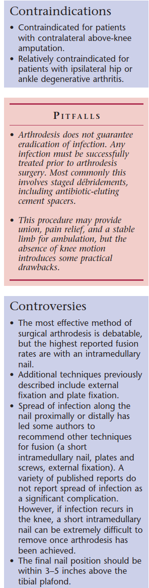
Contraindications
While arthrodesis is a salvage procedure, certain conditions may preclude its successful execution or lead to unfavorable outcomes:
- Active, uncontrolled infection: Although infection is a primary indication, arthrodesis should ideally be performed after infection eradication has been attempted and largely controlled, often after a period with an antibiotic spacer. Performing definitive arthrodesis in the face of rampant, uncontrolled infection risks nonunion and persistent sepsis.
- Insufficient bone stock: Extremely poor bone quality or massive bone defects that prevent stable fixation of the intramedullary nail in both the femur and tibia.
- Severe vascular compromise: Compromised distal perfusion or significant peripheral vascular disease may impair wound healing and bone union, increasing the risk of complications and even amputation.
- Significant ipsilateral hip or ankle pathology: Pre-existing, symptomatic arthritis of the hip or ankle can be exacerbated by the altered biomechanics of a fused knee, leading to progressive pain and disability.
- Patient non-compliance or unwillingness: A fused knee significantly alters gait and lifestyle. Patients must be fully informed and accept these changes and adhere to post-operative protocols.
- Severe soft tissue deficit: Compromised soft tissue envelope, extensive scarring, or large skin defects may complicate wound closure and increase infection risk, potentially requiring plastic surgery consultation for flap coverage.
Operative vs. Non-Operative Indications
The decision matrix for intramedullary arthrodesis is complex, weighing the severity of TKA failure against patient factors and alternative treatments.
| Category | Indication for Intramedullary Arthrodesis (Operative) | Scenarios Where Alternative Surgical or Non-Operative Strategies are Preferred/Attempted First |
|---|---|---|
| Infection | Recalcitrant PJI after multiple failed debridements and reimplantations, especially with severe bone loss. | Acute PJI (debridement and retention or single/two-stage revision); chronic PJI with good bone stock (two-stage revision); non-operative management for mild, well-controlled PJI in frail patients. |
| Extensor Mechanism | Irreparable loss of extensor mechanism (quadriceps/patellar tendon rupture, severe patellar bone loss) causing flail knee. | Primary repair of extensor mechanism ruptures; allograft reconstruction; extensor mechanism plication or advancement for mild insufficiency. |
| Bone Loss | Massive, unreconstructable bone defects after multiple revisions or trauma, preventing stable prosthetic fixation. | Minor to moderate bone loss (modular revision components, augments, cones, allograft reconstruction with revision TKA). |
| Pain/Instability | Persistent, debilitating pain and instability after multiple failed revision TKAs. | Revision TKA for aseptic loosening, instability; pain management for well-aligned, stable TKAs with unclear etiology of pain. |
| Deformity | Severe, rigid deformity that cannot be corrected via revision arthroplasty. | Correctable deformity amenable to revision TKA with appropriate releases and implants. |
| Patient Factors | Young, active patient requiring a durable, stable limb (considering amputation as the only other option). | Elderly, low-demand patient with stable but painful TKA (non-operative management, pain blocks); patients unwilling to accept a fused knee. |
| Contraindications | (Not applicable - these are reasons not to do arthrodesis) | Active, uncontrolled infection; insufficient host bone for nail fixation; severe vascular disease; ipsilateral painful hip/ankle arthritis. |
Pre-Operative Planning & Patient Positioning
Meticulous pre-operative planning is the cornerstone of successful knee arthrodesis, particularly given the complexity of the pathology and the definitive nature of the procedure.
Infection Management
For cases involving PJI, which constitute a large proportion of arthrodesis indications, the infection must be rigorously managed and ideally eradicated
prior
to definitive arthrodesis. This typically involves:
* Explantation of all prosthetic components and cement debris.
* Thorough debridement of necrotic and infected tissues.
* Implantation of an antibiotic-loaded cement spacer.
* Prolonged systemic antibiotic therapy tailored to culture sensitivities.
* Serial inflammatory markers (ESR, CRP) and synovial fluid analysis (if possible) to confirm infection quiescence. A two-stage approach is preferred to optimize infection control before definitive fusion.
Clinical Assessment
- History: Detailed review of prior surgeries, infections, antibiotic regimens, functional limitations, pain characteristics, and patient expectations.
- Physical Examination: Assess hip and ankle range of motion and pain, neurovascular status of the extremity, soft tissue envelope integrity, presence of draining sinuses, and overall limb alignment. Evaluate for any pre-existing LLD.
Imaging
Comprehensive imaging is essential for templating, assessing bone loss, canal morphology, and confirming infection status.
*
Plain Radiographs:
Anteroposterior (AP) and lateral plain films of the affected knee are mandatory. Crucially,
full-length standing AP and lateral radiographs of the entire lower extremity from hip to ankle
are indispensable.
* These films allow for accurate assessment of overall limb alignment, identification of any pre-existing bowing or deformity in the femur or tibia, and determination of the appropriate fusion angle.
* Radiographic magnification markers should be used to enable accurate templating of nail length and diameter.
* They provide information on bone stock, potential bone defects, and any remaining hardware.

* The length of the femur and the tibia must be determined, and any unusual bowing or canal abnormalities must be identified. The length of the femur and the tibia can be obtained from plain radiographs with a measuring template in place.
*
Computed Tomography (CT) Scan:
* A computed tomography (CT) scan from the tip of the trochanter to the distal femur and then from the proximal tibia to the distal tibia can be used. This provides detailed information regarding the intramedullary canal dimensions, particularly identifying the narrowest portion or isthmus of the tibia and femur, which limits the maximum size of the intramedullary nail.
* CT scans are invaluable for identifying any retained cement, previous screw holes that could interfere with new locking screws, areas of osteolysis, or complex bone defects.
* 3D reconstructions can be particularly helpful for visualizing complex deformities and planning reaming trajectories.
*

*
MRI:
Less commonly used due to artifact from retained metal, but can provide insights into soft tissue integrity and occult abscesses if infection is equivocal and CT is non-diagnostic.
*
Nuclear Medicine (e.g., Technetium/Indium-WBC scans):
Utilized when infection status remains uncertain despite clinical and laboratory evaluation.
Nail Templating
Based on comprehensive imaging, the following must be precisely templated:
*
Nail Diameter:
Determined by the narrowest isthmus of both the femoral and tibial canals, often the tibial isthmus. Maximize nail diameter for optimal stiffness.
*
Nail Length:
The arthrodesis nail should extend from the tip of the greater trochanter to well within the isthmus of the distal tibia. This ensures adequate fixation in both segments and bypasses the knee fusion site with sufficient length. Custom-length or custom-bent nails may be required for complex anatomy or significant bowing.
*
Locking Screw Placement:
Plan for a minimum of two, preferably three, locking screws in both the proximal femur and distal tibia. Avoid areas of prior screw holes or significant bone loss.
*
Limb Length:
Pre-operative planning should aim to achieve an acceptable limb length. Significant bone resection may lead to shortening, which must be discussed with the patient and potentially managed with contralateral shoe lifts.
*
Bone Grafting:
Identify areas of anticipated bone loss that will require autograft (e.g., reamings, iliac crest) or allograft supplementation to promote fusion.
Patient Positioning
- Supine Position: The patient is typically positioned supine on a radiolucent operating table.
- Pelvic Support: A small bump under the ipsilateral hip may facilitate access to the greater trochanter.
- Leg Preparation: The entire lower extremity, from the iliac crest to the foot, is prepped and draped to allow full visualization and mobility for accurate nail insertion and image intensifier positioning.
- Image Intensifier: Ensure unrestricted access for both AP and lateral fluoroscopic views of the hip, knee, and ankle throughout the procedure. The C-arm should be draped sterilely.
- Tourniquet: A high thigh tourniquet may be used to minimize blood loss, but its use should be weighed against the patient's vascular status, especially in infected cases where reperfusion for antibiotic delivery is beneficial.
Detailed Surgical Approach / Technique
The execution of intramedullary arthrodesis requires meticulous surgical technique, especially given the altered anatomy and potential for persistent infection.
1. Preparation and Incision
- Pre-operative antibiotics: Administer standard prophylactic antibiotics (and potentially organism-specific antibiotics for known infection) prior to incision.
- Sterile Prep and Drape: The limb is prepped from the iliac crest to the toes. A sterile impermeable drape isolating the foot from the rest of the surgical field is often utilized.
- Incision: A standard midline anterior incision over the knee is generally preferred. If previous incisions are present, utilize them if feasible, extending proximally over the distal femur and distally over the proximal tibia as necessary to adequately expose the bone and facilitate intramedullary access. For femoral nailing, a separate small incision over the greater trochanter may be required.
2. Exposure and Debridement
- Joint Exposure: Develop skin and subcutaneous flaps to expose the distal femur, knee joint space (or what remains of it), and proximal tibia.
- Explantation and Debridement: All remaining components of the failed TKA, including cement, polyethylene, and metal implants, must be meticulously removed. This is often the most challenging part of the procedure, especially with well-fixed components or extensive cement mantle.
- Infection Control: In cases of infection, perform thorough synovectomy and debridement of all granulation tissue, necrotic bone, and fibrous membranes. Send multiple tissue samples for aerobic, anaerobic, fungal, and atypical mycobacterial cultures. Pulsed lavage is used liberally.
- Extensor Mechanism: Address the extensor mechanism. Often, the patella is excised (patellectomy) if compromised or extensively damaged, simplifying the fusion. If a functional extensor mechanism remains, it should be preserved if possible, but often the knee capsule is opened wide to gain access.
3. Joint Preparation and Resection
-
Bone Preparation:
The goal is to create broad, bleeding cancellous bone surfaces on the distal femur and proximal tibia that will achieve union.
- Ream any sclerotic bone or retained cement from the distal femur and proximal tibia.
- Use oscillating saws or osteotomes to prepare flat or slightly contoured bone surfaces on both the femur and tibia. The amount of bone resection should be minimized to preserve limb length while ensuring good contact.
- Consider creating a tongue-in-groove or stepped resection to enhance rotational stability.
-

- Fusion Angle: Position the knee in the desired fusion angle (typically 0-10 degrees of flexion, neutral varus/valgus, and slight external rotation). This can be achieved by angling the resection cuts or by manipulating the limb during nail insertion.
- Bone Grafting: Autogenous bone graft (harvested from reamings, iliac crest, or proximal tibia) should be packed into any remaining defects at the fusion site to promote osteogenesis and enhance union rates. Allograft chips or synthetic bone graft substitutes can be used to augment.
4. Intramedullary Nail Insertion
-
Femoral Entry Point:
- Proximal Femur: A separate incision, or an extension of the primary incision, is made over the tip of the greater trochanter. A starting awl or drill is used to create an entry point just medial to the tip of the greater trochanter, in line with the femoral shaft axis. This entry point is crucial to avoid varus malalignment.
- Guide Wire Insertion: A long guide wire is inserted down the femoral canal, passing through the resected knee joint space, and into the proximal tibia. Fluoroscopy (AP and lateral views) is essential to confirm correct guide wire placement in the center of both femoral and tibial canals.
-

-
Reaming:
- Sequential reaming begins in the femur, following the guide wire, to the predetermined diameter and depth. This clears the canal and provides cancellous bone for grafting.
- The guide wire is then advanced further into the tibia, and sequential reaming of the tibia is performed to the desired diameter and length. Ensure reamers pass smoothly across the fusion site. The largest possible diameter nail, compatible with the narrowest isthmus, should be used.
-
Nail Insertion:
- The chosen intramedullary nail, typically a custom-length, custom-diameter arthrodesis nail designed for unicompartmental knee fusion (long segment nail), is carefully advanced over the guide wire.
- The nail should pass across the fusion site smoothly, achieving firm engagement in both the femur and tibia.
- Confirm the final position and alignment with fluoroscopy. The nail should extend from the greater trochanter to the distal tibial metaphysis, bypassing the fusion site completely.
-

5. Locking and Compression
- Distal Locking (Tibia): Once the nail is in optimal position, distal locking screws are inserted through the nail using a targeting guide. A minimum of two, and preferably three, interlocking screws are placed in the distal tibia. Confirm screw length and position with AP and lateral fluoroscopy.
- Proximal Locking (Femur): Proximal locking screws are then inserted into the femur, also using a targeting guide. A minimum of two, and preferably three, screws are placed in the proximal femur, typically through the greater trochanter incision. Again, confirm with fluoroscopy.
-
Compression:
If there is any distraction at the fusion site, or if the nail design allows, compression can be applied through the nail to further promote bony apposition and healing. This may involve specific nail designs with compression bolts or dynamic locking options.


6. Wound Closure
- Drains: Consider placing a negative pressure drain in the joint space to manage hematoma and reduce dead space, especially in infected cases.
- Layered Closure: Close the surgical incision in layers, ensuring meticulous hemostasis and tension-free skin closure.
- Dressings: Apply sterile dressings and potentially a soft compressive bandage.
Complications & Management
Despite its effectiveness as a salvage procedure, intramedullary knee arthrodesis is associated with a significant complication profile, reflecting the complexity of the underlying pathology and the extensive surgical intervention.
Common Complications and Salvage Strategies
| Complication | Incidence (Approximate) | Description & Risk Factors | Salvage Strategies |
|---|---|---|---|
| Infection Recurrence | 10-25% | Persistence or recurrence of periprosthetic joint infection (PJI), especially common in cases where arthrodesis was performed for PJI. Risk factors include virulent organisms, host compromise, inadequate debridement, and severe soft tissue damage. | Extensive surgical debridement with implant retention (if stable and infection controlled early), implant removal with antibiotic-loaded cement spacer followed by revision arthrodesis or amputation. Long-term suppressive antibiotic therapy. |
| Nonunion/Delayed Union | 10-30% | Failure of bony fusion, often defined as no radiographic evidence of union by 6-9 months post-op. Risk factors: persistent infection, smoking, poor bone stock, inadequate fixation, large bone gaps, revision surgery. | Revision surgery with repeat debridement, aggressive bone grafting (autograft preferred, e.g., iliac crest or reamings), potential exchange nailing to a larger diameter or stiffer implant, compression across the fusion site. Adjunctive therapies: electrical stimulation, pulsed electromagnetic fields (PEMF), low-intensity pulsed ultrasound (LIPUS). |
| Malunion | 5-15% | Fusion in an undesirable position (e.g., excessive flexion, varus/valgus, or rotation). Impacts gait, sitting, and adjacent joint stress. Risk factors: inadequate intraoperative fluoroscopy, poor surgical technique, severe pre-existing deformity. | Minor malunion: shoe modifications. Severe malunion: corrective osteotomy at the fusion site, potentially with revision nailing and repeat fixation. May require custom-bent nails or external fixation for temporary correction. |
| Hardware Failure | 5-10% | Nail breakage, screw loosening, or migration. Often a consequence of nonunion or prolonged unprotected weight-bearing. Risk factors: poor bone quality, early weight-bearing, persistent nonunion, stress shielding. | Revision surgery with implant removal and exchange nailing (often to a larger, stiffer implant). Address underlying nonunion with bone grafting and compression. May require supplementary plate fixation if bone quality is poor or nail exchange is not feasible. |
| Neurovascular Injury | <5% | Damage to the popliteal artery/vein or tibial/peroneal nerves during reaming, debridement, or screw placement. Risk factors: extensive scarring, anatomical variations, aggressive dissection. | Immediate vascular repair for arterial injury. Nerve repair or neurolysis for significant nerve injury. Post-operative observation and physical therapy for neuropraxia. Prevention is key through careful dissection and anatomical awareness. |
| Limb Length Discrepancy (LLD) | 10-20% | Shortening of the affected limb due to bone resection or lack of distraction during fusion. Risk factors: extensive bone loss requiring significant resection, technical error. | Minor LLD (<2cm): shoe lift or orthotic. Moderate LLD (2-5cm): shoe lift, consideration for contralateral limb lengthening (rare). Severe LLD (>5cm): complex reconstructive limb lengthening (very rare in this context). |
| Adjacent Joint Arthrosis | Long-term complication | Accelerated degenerative changes in the ipsilateral hip or ankle due to altered biomechanics and increased stress. Risk factors: pre-existing arthritis, improper fusion angle, active lifestyle. | Conservative management initially (NSAIDs, activity modification, physical therapy). Intra-articular injections. Eventual arthroplasty of the hip or ankle if symptoms become debilitating and conservative measures fail. |
| Hematoma/Seroma | 5-10% | Collection of blood or serous fluid in the wound, increasing infection risk. Risk factors: inadequate hemostasis, poor drainage, extensive dissection. | Aspiration, drain insertion, or surgical evacuation. Close monitoring for signs of infection. |
| Wound Healing Issues | 5-15% | Superficial or deep wound dehiscence, skin necrosis, or fistula formation. Risk factors: poor soft tissue quality, previous incisions, infection, tension on closure. | Local wound care, débridement, negative pressure wound therapy (NPWT). May require plastic surgery consultation for local or free flap coverage in severe cases. |
| DVT/PE | 1-5% (with prophylaxis) | Deep vein thrombosis or pulmonary embolism. Risk factors: prolonged immobilization, extensive surgery, obesity, hypercoagulable states. | Standard pharmacological prophylaxis (anticoagulants). Mechanical prophylaxis (intermittent pneumatic compression). Early mobilization post-operatively. Treatment with therapeutic anticoagulation for confirmed events. |
Post-Operative Rehabilitation Protocols
Post-operative rehabilitation following knee arthrodesis is designed to protect the fusion site, promote bone healing, manage pain, and restore maximal functional independence. The primary goal is achieving solid fusion, making early uncontrolled motion detrimental.
Early Post-Operative Phase (Days 0-6 Weeks)
- Pain Management: Multimodal analgesia including regional blocks, NSAIDs (if no contraindication), acetaminophen, and opioids as needed. Adequate pain control facilitates early mobilization.
- Wound Care: Daily dressing changes, monitoring for signs of infection, hematoma, or dehiscence. Drain removal typically within 24-72 hours when output is minimal.
- Deep Vein Thrombosis (DVT) Prophylaxis: Standard pharmacological (e.g., low molecular weight heparin) and mechanical (e.g., intermittent pneumatic compression devices) prophylaxis should be initiated immediately and continued according to institutional guidelines, often for 4-6 weeks.
-
Weight-Bearing:
This is a critical decision and depends on the stability of the fixation, quality of bone, and surgeon preference.
- Initial: Typically non-weight-bearing (NWB) or touch-down weight-bearing (TDWB) with crutches or a walker to protect the fusion site.
- Progression: Gradual progression to partial weight-bearing (PWB) may begin as early as 2-4 weeks post-operatively if fixation is very stable, pain is controlled, and radiographs show no issues.
-
Mobility:
- Bed Mobility: Early active hip and ankle range of motion (ROM) exercises to prevent stiffness and improve circulation.
- Transfers: Practice safe transfers from bed to chair.
- Gait Training: With the assistance of physical therapy, initiate gait training with NWB or TDWB using appropriate assistive devices. Education on compensatory gait patterns with a fused knee.
- Edema Management: Elevation of the limb, compression stockings, and ankle pumps to reduce swelling.
Intermediate Phase (6 Weeks - 3 Months)
- Radiographic Assessment: Obtain serial radiographs (AP and lateral) at 6 weeks, 3 months, and 6 months to monitor for signs of union (callus formation, trabecular bridging) and assess hardware integrity.
-
Weight-Bearing Progression:
- As radiographic signs of healing progress and pain subsides, weight-bearing can be gradually increased from PWB to weight-bearing as tolerated (WBAT) .
- Wean off assistive devices as strength, balance, and confidence improve.
-
Strengthening:
- Focus on strengthening of the hip abductors, adductors, flexors, and extensors.
- Ankle dorsiflexors and plantarflexors strengthening to compensate for altered gait mechanics.
- Core stability exercises.
- Gait Training: Continue intensive gait training to optimize efficiency and minimize compensatory movements. Address any limping or awkwardness.
- Proprioception and Balance: Initiate balance exercises as weight-bearing progresses.
Late Phase (3 Months - 6 Months and Beyond)
- Radiographic Confirmation: Full weight-bearing is typically allowed once radiographic evidence of solid bony union is confirmed (often by 4-6 months, but can take longer, up to 9-12 months).
- Full Weight-Bearing: Progress to full, unassisted weight-bearing.
-
Advanced Strengthening and Conditioning:
- Continue a comprehensive home exercise program focused on lower extremity and core strength.
- Low-impact cardiovascular activities (e.g., swimming, cycling with one leg) can be incorporated.
- Activity Modification: Educate the patient on activity modifications for a fused knee. High-impact activities or prolonged standing may be difficult.
- Limb Length Discrepancy Management: If significant LLD is present, assess for shoe lifts or orthotics.
- Long-Term Monitoring: Periodic follow-up to monitor for adjacent joint issues (hip, ankle) and ensure long-term functional independence. Patients should be counseled on the potential for secondary arthrosis in these joints.
Summary of Key Literature / Guidelines
Intramedullary knee arthrodesis for failed TKA, while a highly effective salvage option, remains a relatively uncommon procedure, meaning large randomized controlled trials are scarce. The evidence base largely comprises case series, retrospective reviews, and expert consensus.
Fusion Rates and Outcomes
- High Fusion Rates: Studies consistently report high fusion rates ranging from 80% to 100% with intramedullary nailing. This is significantly higher than historical external fixation methods. Factors influencing fusion include meticulous surgical technique, adequate debridement of infected tissue, robust fixation, and bone grafting.
- Pain Relief: The primary goal of arthrodesis is to achieve a pain-free, stable limb. Most patients report significant pain relief post-fusion, contributing to improved quality of life despite the loss of knee motion.
- Functional Outcomes: While knee motion is eliminated, functional outcomes are generally favorable for patients who would otherwise face amputation or a flail, painful limb. Patients can ambulate, stand, and participate in many activities of daily living. Gait is altered, often characterized by a stiff-legged swing phase and increased demands on hip and ankle.
- Complications: As detailed above, complications remain significant. Infection recurrence, nonunion, and malunion are the most frequently cited. The cumulative reoperation rate can be substantial, emphasizing the challenging patient population and complex nature of the procedure.
Key Literature and Consensus
- Biomechanical Superiority of IM Nailing: Early comparative studies established the biomechanical superiority of intramedullary nails over external fixation or plate constructs for arthrodesis, demonstrating increased stiffness and load sharing, which translated into higher fusion rates and reduced hardware complications.
- Management of PJI: Consensus guidelines for periprosthetic joint infection, such as those from the International Consensus Group on Periprosthetic Joint Infection (ICM PJI), acknowledge knee arthrodesis as a viable and sometimes necessary option for definitive infection control in recalcitrant cases, often in a staged protocol following explantation and antibiotic spacer placement.
- Optimal Fusion Position: While slight variations exist, the general consensus recommends a few degrees of flexion (0-10°) and neutral varus/valgus for optimal functional outcomes, balancing stability with swing-phase clearance and sitting comfort.
- Role of Bone Grafting: The literature supports the use of autogenous bone graft (e.g., reamings, iliac crest) to enhance fusion rates, especially in cases with significant bone loss or revision surgery where the biological environment for healing may be compromised. Allograft can be used as an extender or filler.
- Custom vs. Universal Nails: While many universal intramedullary nails can be adapted, custom-length and custom-bent arthrodesis nails (often unlinked femoral-tibial nails designed specifically for this purpose) are frequently preferred to accommodate varied anatomy, bypass bone defects, and achieve optimal fixation length.
- Multidisciplinary Approach: The management of these complex patients often benefits from a multidisciplinary team including orthopedic surgeons, infectious disease specialists (for PJI), pain management specialists, and physical therapists.
Future Directions
Ongoing research focuses on optimizing surgical techniques, improving implant designs for even greater stability, and exploring biological adjuncts to enhance fusion (e.g., growth factors, mesenchymal stem cells). Additionally, long-term studies on adjacent joint kinematics and arthritis progression will continue to refine our understanding of this salvage procedure. While certainly not a first-line treatment, intramedullary arthrodesis remains an invaluable tool in the armamentarium of the orthopedic surgeon for solving the most challenging cases of failed total knee arthroplasty.
Clinical & Radiographic Imaging

