Introduction to Os Acromiale and Impingement Syndrome
The acromion plays a pivotal biomechanical role in shoulder function, serving as the origin for the middle and anterior deltoid and providing the superior structural boundary of the subacromial space. An os acromiale represents a failure of fusion of one or more of the acromial ossification centers. While frequently an incidental radiographic finding, an unstable os acromiale can become a significant source of shoulder pathology, specifically contributing to dynamic subacromial impingement syndrome (SAIS) and, in chronic cases, rotator cuff tears.
Understanding the embryology, pathoanatomy, and precise surgical indications is paramount for the orthopedic surgeon. Misdiagnosis or improper surgical execution—such as aggressive resection leading to deltoid detachment—can result in catastrophic functional deficits. This guide provides an exhaustive, evidence-based approach to the evaluation and surgical management of os acromiale in the setting of impingement.
Embryology and Pathoanatomy
The Four Ossification Centers
The acromion develops from four distinct ossification centers. These centers typically appear around age 15 and normally fuse to one another by age 18. The fully formed acromion then fuses to the scapular spine between the ages of 20 and 25 years. Failure of fusion at any of these junctions results in an os acromiale.
The four ossification centers, from anterior to posterior, are:
1. Preacromion: The most anterior tip of the acromion.
2. Mesoacromion: The middle segment, which forms the bulk of the lateral acromion.
3. Metaacromion: The posterior segment, located near the acromial angle.
4. Basiacromion: The base of the acromion, attaching directly to the scapular spine.
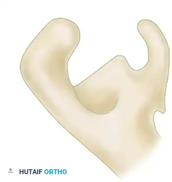
Clinical Pearl: The most common site for nonunion (and thus the most common location for an os acromiale) lies between the mesoacromion and metaacromion.
Epidemiology
Os acromiale is present in approximately 2.7% of the general population. A critical diagnostic factor for the orthopedic surgeon to remember is its high rate of bilaterality; it is bilateral in 60% of individuals who possess the anomaly. Therefore, if an os acromiale is identified on the symptomatic shoulder, the contralateral shoulder should be evaluated, even if asymptomatic, to establish a baseline.
Biomechanics of Dynamic Impingement
Most instances of os acromiale are entirely asymptomatic. However, when symptoms do arise, they are typically driven by a dynamic biomechanical conflict.
The deltoid muscle originates from the lateral and anterior margins of the acromion. In the presence of a fibrous nonunion (os acromiale), the contraction of the deltoid during active shoulder abduction and forward elevation exerts a downward pull on the unfused anterior fragment (usually the mesoacromion or preacromion). This creates a "hinge effect," causing the unfused fragment to tilt inferiorly into the subacromial space. This dynamic depression directly impinges upon the supraspinatus tendon and the subacromial bursa, leading to bursitis, tendinopathy, and potentially rotator cuff tearing over time.
Clinical Evaluation and Evidence-Based Literature
The Wolf Study on Arthroscopic Management
The association between os acromiale and impingement syndrome has been a subject of debate. In a landmark study of 294 consecutive patients treated with arthroscopic techniques for impingement, Wolf identified an os acromiale in 3.1% of the cohort.
Key findings from this study include:
* Prevalence: The 3.1% prevalence in the impingement group was not considered indicative of an increased prevalence of impingement over the general population (which sits at ~2.7%).
* Rotator Cuff Integrity: Only one patient with an os acromiale in this series had a complete rotator cuff tear, suggesting that while it causes impingement, it may not rapidly accelerate full-thickness cuff failure compared to standard Type III hooked acromions.
* Surgical Success: Nine patients underwent arthroscopic resection of the os acromiale using a burr. A critical technical note from Wolf was the strict preservation of the deltopectoral fascia.
* Outcomes: Eight of the nine operations were rated as successful based on the UCLA shoulder rating scale.
Surgical Warning: The primary mechanism of failure in os acromiale surgery is iatrogenic deltoid detachment. Wolf’s success was predicated on preserving the superior fibrous envelope (the deltoid aponeurosis) while resecting the bony fragment from the undersurface.
Diagnostic Imaging Protocol
A meticulous radiographic evaluation is required to differentiate an os acromiale from a normal acromioclavicular joint, a fracture, or a standard acromial spur. We prefer to evaluate impingement with a standard, high-quality radiograph series.
1. Standard Radiographic Series
- Anteroposterior (AP) View: Often unhelpful for diagnosing os acromiale due to the overlapping shadows of the clavicle and the scapular spine. However, it is essential for evaluating glenohumeral arthritis and superior humeral head migration.
- Supraspinatus Outlet View (Y-View): Excellent for assessing acromial morphology (Biggliani classification) and identifying subacromial spurs. The os acromiale can frequently be visualized here as a double-density or a distinct cleft in the acromial arch.
- Axillary Lateral View: The gold standard for diagnosing os acromiale. This view profiles the acromion from anterior to posterior, clearly delineating the radiolucent line of the nonunion between the ossification centers.
2. Advanced Imaging (MRI and CT)
- Magnetic Resonance Imaging (MRI): Highly recommended for surgical planning. MRI evaluates the integrity of the rotator cuff and the subacromial bursa. Crucially, fluid-sensitive sequences (T2/STIR) will demonstrate bone marrow edema across the synchondrosis of the os acromiale. The presence of edema strongly correlates with instability and a symptomatic os acromiale, differentiating it from an incidental, asymptomatic finding.
- Computed Tomography (CT): Useful for preoperative planning if Open Reduction and Internal Fixation (ORIF) is being considered for a large mesoacromion fragment, as it provides excellent bony detail of the fragment size and the nonunion interface.
Surgical Indications and Decision-Making
Surgical intervention is indicated only after a comprehensive trial of conservative management (minimum 3 to 6 months), which should include NSAIDs, physical therapy focusing on rotator cuff and periscapular stabilization, and subacromial corticosteroid injections.
The surgical approach is dictated by the anatomical location of the nonunion and the thickness of the acromion.
Indication 1: Mesoacromion-Metaacromion Nonunion
If the os acromiale is located between the mesoacromion and metaacromion (the most common site) and the acromion is of normal thickness:
* Procedure of Choice: Arthroscopic undersurface acromioplasty.
* Rationale: Resecting the undersurface to make it flat eliminates the dynamic impingement without compromising the superior deltoid attachment or requiring complex osteosynthesis.
Indication 2: Preacromion-Mesoacromion Nonunion
If an os acromiale is present between the preacromion and mesoacromion (a smaller, more anterior fragment):
* Procedure of Choice: Complete arthroscopic or open resection of the preacromion fragment.
* Rationale: The preacromion is small enough that its complete removal does not significantly compromise the deltoid origin, provided the fibrous attachment of the deltoid and trapezius is meticulously left intact and repaired side-to-side.
Indication 3: Large, Unstable Mesoacromion (Alternative Approach)
While undersurface resection is highly successful, some authors advocate for ORIF (bone grafting and tension band wiring or screw fixation) for very large, highly unstable mesoacromial fragments in young, high-demand laborers to preserve the deltoid lever arm. However, this carries a higher risk of nonunion and hardware irritation.
Step-by-Step Surgical Technique: Arthroscopic Management
The following details the arthroscopic undersurface resection and fragment excision, which remains the workhorse procedure for symptomatic os acromiale.
1. Patient Positioning and Anesthesia
- Anesthesia: General anesthesia supplemented with an interscalene regional nerve block for postoperative pain control.
- Positioning: The patient can be placed in either the beach-chair or lateral decubitus position, based on surgeon preference. The beach-chair position allows for easier conversion to an open procedure if deltoid repair becomes necessary.
- Preparation: Standard sterile prep and drape, ensuring the arm is freely mobile.
2. Portal Placement and Diagnostic Arthroscopy
- Establish a standard posterior viewing portal.
- Perform a thorough diagnostic glenohumeral arthroscopy to rule out intra-articular pathology (e.g., SLAP tears, articular-sided rotator cuff tears).
- Enter the subacromial space. Establish a standard lateral working portal and an anterior portal.
3. Subacromial Preparation
- Use a radiofrequency wand and an arthroscopic shaver to perform a thorough subacromial bursectomy.
- Clear the soft tissue from the undersurface of the acromion to clearly visualize the coracoacromial (CA) ligament and the os acromiale synchondrosis.
- Identification: The nonunion site will often appear as a fibrous cleft or a step-off on the undersurface of the acromion. Probing the anterior fragment will often demonstrate gross instability.
4. Technique A: Undersurface Acromioplasty (For Meso-Meta Nonunion)
- Introduce a high-speed arthroscopic burr through the lateral portal.
- Begin resecting the undersurface of the anterior fragment (the mesoacromion).
- The goal is to thin the fragment and create a flat, smooth transition from the anterior acromion to the posterior acromion, eliminating the impingement spur.
- Crucial Step: Do not resect through the superior cortex. The superior periosteal envelope and the deltoid aponeurosis must remain completely intact. Resecting too aggressively superiorly will detach the deltoid.
5. Technique B: Complete Fragment Resection (For Preacromion Nonunion)
- If the fragment is a small preacromion, it can be completely excised.
- Use the burr to carefully shell out the bony fragment from within its soft tissue envelope.
- Work from the undersurface superiorly. As the bone is thinned to an eggshell, use a curette or the burr to gently remove the remaining bone, leaving the superior fibrous tissue (the confluence of the trapezius and deltoid fascia) completely intact.
- If the CA ligament is attached to the resected fragment, it is released during this process, which further decompresses the subacromial space.
Pitfall: Failure to recognize a large mesoacromion and attempting complete excision rather than undersurface acromioplasty will result in a massive deltoid defect. Always assess fragment size on the preoperative axillary lateral radiograph.
6. Final Assessment and Closure
- Take the arm through a dynamic range of motion while viewing from the posterior portal to ensure that the impingement has been completely eliminated and that the supraspinatus tendon glides smoothly beneath the preserved deltoid fascia.
- Thoroughly irrigate the subacromial space to remove all bone debris, which can cause postoperative heterotopic ossification.
- Close the portals with non-absorbable sutures.
Postoperative Rehabilitation Protocol
The postoperative protocol must balance the need to prevent stiffness with the absolute necessity of protecting the deltoid origin, especially if the bone was thinned significantly or a small fragment was excised.
Phase I: Protection (Weeks 0-4)
- Immobilization: The patient is placed in a standard shoulder sling.
- Range of Motion: Passive range of motion (PROM) is initiated immediately to prevent adhesive capsulitis. Forward elevation and external rotation are permitted within the limits of pain.
- Restrictions: Strict avoidance of active shoulder abduction and forward elevation. Active contraction of the deltoid places stress on the thinned acromion or the preserved fascial envelope, risking dehiscence.
Phase II: Active Motion (Weeks 4-8)
- Sling: Discontinue the sling.
- Range of Motion: Transition from active-assisted range of motion (AAROM) to active range of motion (AROM).
- Strengthening: Begin gentle, closed-chain isometric deltoid and rotator cuff strengthening. Focus heavily on periscapular stabilizers (rhomboids, serratus anterior) to optimize scapulothoracic rhythm.
Phase III: Advanced Strengthening (Weeks 8-12+)
- Progress to isotonic strengthening of the rotator cuff and deltoid.
- Incorporate plyometrics and sport-specific or work-specific functional training.
- Full return to heavy labor or overhead sports is typically permitted between 3 and 5 months postoperatively, provided the patient has full, painless ROM and symmetric strength.
Complications and Management
While arthroscopic management of os acromiale is highly successful (as evidenced by Wolf's 89% success rate), complications can occur:
- Deltoid Dehiscence: The most devastating complication. Caused by over-resection of the superior cortex and violation of the deltoid aponeurosis. Presents with profound weakness in abduction and a palpable defect. Requires complex open surgical repair, often utilizing tissue grafts.
- Inadequate Resection: Failure to remove enough of the undersurface, leading to persistent dynamic impingement and refractory pain. Requires revision arthroscopy.
- Heterotopic Ossification: Bone debris left in the subacromial space can ossify, causing recurrent impingement. Meticulous intraoperative irrigation is preventative.
- Acromial Fracture: Aggressive thinning of a large mesoacromion can leave the remaining bone structurally weak, leading to a stress fracture.
Conclusion
Os acromiale is a critical differential diagnosis in the evaluation of subacromial impingement syndrome. A thorough understanding of acromial embryology and the biomechanics of dynamic impingement is essential. By utilizing the axillary lateral radiograph and MRI, surgeons can accurately diagnose a symptomatic os acromiale. When conservative measures fail, arthroscopic undersurface acromioplasty or targeted fragment excision—with uncompromising preservation of the deltopectoral fascia—provides excellent, reproducible clinical outcomes for the orthopedic patient.
📚 Medical References
- os acromiale, J Am Acad Orthop Surg 14:12, 2006.
- Lapidus PW: Infi ltration therapy of acute bursitis with calcium, Surg Gynecol Obstet 76:715, 1943.
- Lee JC, Sykes C, Saifuddin A, et al: Adhesive capsulitis: sonographic changes in the rotator cuff interval with arthroscopic correlation, Skeletal Radiol 34:522, 2005.
- Leffert RD: The frozen shoulder, Instr Course Lect 34:199, 1985.
- Lindholm K: On pathogenesis of ruptures of the tendon aponeurosis of the shoulder joint, Acta Radiol 20:563, 1939.
- Linker CS, Helms CA, Fritz RC: Quadrilateral space syndrome: fi ndings at MR imaging, Radiology 188:675, 1993.
- Lippmann RK: Frozen shoulder, periarthritis, bicipital tenosynovitis, Arch Surg 47:283, 1943.
- Liu SH, Baker CL: Arthroscopically assisted rotator cuff repair: correlation of functional results with integrity of the cuff, Arthroscopy 10:54, 1994.
- Lo IKY, Bishop JY, Miniaci A, et al:
