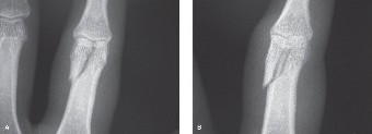
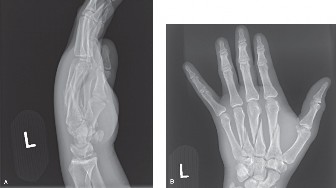
Orthopedic Hand Cases: A Patient's Journey, Is a Year Old a Factor?
Updated: Feb 2026
69 Views
Key Medical Takeaway
For anyone wondering about Orthopedic Hand Cases: A Patient’s Journey, Is a Year Old a Factor?, For multiple displaced metacarpal fractures, open reduction and internal fixation (ORIF) is the most appropriate management, particularly in high-energy trauma. When the patient is a 28-year-old male, ORIF restores anatomy, including longitudinal and transverse arches, decompresses interosseous muscles, and stabilizes the hand. This enables earlier rehabilitation, significantly optimizing the patient’s functional outcome.
Clinical Radiograph / Orthopedic Image Figure 4–1 A–B
He was then splinted and presented to your office on the fourth day after injury.
The most appropriate management at this time for this injury would be:
Short-arm splint for 6 weeks
Short-arm cast in intrinsic plus position for 6 weeks
Long-arm cast in intrinsic plus position for 6 weeks
Open reduction, internal fixation of all fractures
Discussion
The correct answer is (D). This patient has suffered multiple displaced metacarpal
fractures in contiguous digits. The most appropriate treatment would be an open reduction and internal fixation in order to give the patient an earlier, rehabilitative start.
Identify sequela from this type of injury?** CASE 11 **A 26-year-old female was traveling with her fiancé. At a rest stop, as she got out of the car, he accidentally shut the car door on her left ring finger. There was immediate swelling and bleeding, and after application of first aid, the finger remained swollen and the patient is now here to see you. Radiographs do not show any obvious bony abnormality.
* **Clinical examination of the finger reveals a swollen finger, a tender pulp, and a subungual hematoma, which occupies approximately 50% of the nail plate. The next step in management would be: [View Source / PubMed]
4. Open repair, release of the eponychial fold, exploration of the sterile and germinal matrix, and replacement of the nail plate as a stent
## _Discussion_
The correct answer is (C). As described, the patient does not have severe discomfort. In the absence of an obvious fracture, the only indication for drainage of a subungual hematoma would be for pain control where the patient has excruciating pain from a subungual hematoma. Since that is not the case, this subungual hematoma does not need to be drained. Traditional teaching has suggested that if a subungual hematoma exceeds 30% to 40% of the size of the nail plate, then the nail should be removed and the nail bed should be repaired. However, longitudinal studies have shown that removal of the nail plate and repair of the nail bed does not appear to influence nail growth positively in most patients. Therefore, in the absence of a fracture and if the nail plate is well fixed, irrespective of the size of the hematoma, not only does the hematoma not need to be drained, but the nail plate should not be removed, and the nail bed does not need repair. In this situation,
the patient’s finger may be splinted for comfort for a few days, elevation and icing is recommended, and range of motion is started at the earliest possibility.
At the same time that the patient injured her ring finger, the middle finger also sustained an injury. The middle finger radiographs, however, show that she has a fracture of the distal phalanx which is essentially nondisplaced and a subungual hematoma which occupies 50% of the size of the nail plate.
* **The most appropriate management for the middle finger would be which of the following? [View Source / PubMed]
4. Splinting for comfort and range of motion to be started as soon as comfortable
## _Discussion_
The correct answer is (D). Although the patient has a hematoma which occupies 50% of the nail plate, the description of this finger suggests that the fracture of the distal phalanx is completely nondisplaced. In such situations, there is no indication to remove the nail plate and repair the nail bed. A well-fixed nail plate and nondisplaced fracture of the distal phalanx essentially form a splint for the nail bed and allow the fracture and nail bed to heal in as anatomical position as possible. By removing a well fixed nail plate, the nail bed is destabilized and the support that the nail plate would afford for distal phalangeal fracture is also lost. Therefore, the nail plate is not to be removed in this situation. the patient would simply benefit from a splint and starting range of motion program as soon as the fracture gets more comfortable which is usually around 2 to 3 weeks.
During the same accident, the patient also sustained a fracture of the index finger. The index finger showed a fracture of the distal phalanx which was displaced. The fracture was at the base, and the nail plate was elevated in the proximal eponychial fold from which there was bleeding.
* **The most appropriate management for this injury would be which of the following? [View Source / PubMed]
1. Removal of the nail plate, replacement of the distal phalanx, reduction of the distal phalangeal fracture, fixation with Kirschner wire, open repair of the nail bed, and repair of the nail plate or stenting open the eponychial fold [View Source / PubMed]
4. Volar approach plate fixation of the distal phalanx
## _Discussion_
The correct answer is (A). This digit has a displaced fracture with proximal avulsion of the nail plate from the eponychial fold. This situation involves an injury to the germinal matrix of the nail bed and one which is unlikely to heal in the most optimal circumstances unless the distal phalanx is repositioned anatomically, fixed, and then the nail bed is repaired meticulously. To do this, the nail plate is initially removed. The laceration and the nail bed are carefully assessed. If necessary, small incisions are made in the lateral eponychial folds so that the eponychial fold can be folded back. After irrigation and debridement of the fracture site, it is reduced and carefully pinned with a wire. Then, the nail bed is repaired carefully with absorbable sutures, usually chromic catgut. The nail plate is cleaned and repositioned in the nail fold to act as a stent to keep it open in order for the new nail to grow back. The patient should be cautioned about unpredictability of nail growth, that nail growth can take 6 months to stabilize, and to understand the final outcome from a nail bed repair.** Objectives: Did you learn...? **Idenftify indications for subungal hematoma drainage? Treat a distal phalanx fracture?** CASE 12 **[A 24-year-old, law student injured her left index finger during a volleyball game and developed immediate pain and deformity. By her description, this deformity occurred at the level of the PIP joint. Courtside, one of her colleagues immediately pulled on the finger, and she presents to your office 3 days later. Clinically, she has a swollen finger, but there is no obvious deformity. Radiographs are shown in ](file:///C:/Users/DELL/Desktop/hip/board/Chapter%204.%20The%20Hand_Converted.html#bookmark10)[Figure ](file:///C:/Users/DELL/Desktop/hip/board/Chapter%204.%20The%20Hand_Converted.html#bookmark10)4–10A [and ](#bookmark10)B.

_**Figure 4–10 A–B**_
* **The most likely diagnosis is which of the following? [View Source / PubMed]
4. Bony avulsion of the flexor digitorum superficialis
## _Discussion_
The correct answer is (A). Radiographs show a unicondylar fracture of the proximal phalanx, in this case involving the ulnar condyle. The condyle is displaced, and there appears to be an articular stepoff. The patient’s description of the injury and deformity are consistent with what could have occurred at the time of the injury, and the displacement at the time of injury may have been more significant than that being noted on the radiographs at this time.
* **The most appropriate form of treatment at this time would be which of the following? [View Source / PubMed]
5. Either C or D
## _Discussion_
The correct answer is (E). This is an unstable injury as evidenced by the patient’s description of the deformity at the time of injury and by the radiographs seen in the office. The fracture is displaced and is an intra-articular fracture. There is an articular stepoff. Furthermore, it is an oblique fracture. All of these features indicate that this is an unstable injury, and treatment by closed means is unlikely to be successful. In fractures that are completely nondisplaced, closed nonoperative
[treatment remains an option. However, in this instance where there is articular stepoff and displacement has occurred, it is vital to restore the length of the fragment, and more critically restore articular congruity especially in this young person. Closed reduction and percutaneous fixation as shown in the postoperative radiographs can be effective if perfect reduction of the joint is achieved (](file:///C:/Users/DELL/Desktop/hip/board/Chapter%204.%20The%20Hand_Converted.html#bookmark11)[Fig. ](file:///C:/Users/DELL/Desktop/hip/board/Chapter%204.%20The%20Hand_Converted.html#bookmark11)4–10C [to ](#bookmark11)E). Failure to achieve perfect joint reduction by closed means necessitates an open reduction and treatment with either pins or screws.


_**Figure 4–10 C–E**_
* **Should open reduction and internal fixation of this joint be performed, which of the following are likely complications? [View Source / PubMed]
The correct answer is (C). Irrespective of which treatment is utilized in this instance, it is almost always safe to allow the patient gentle range of motion of the DIP joint. Therefore, stiffness of the DIP joint is usually not a concern in these injuries. However, during open reduction internal fixation of this injury it is vital to maintain the attachment of the collateral ligament to the fractured proximal phalanx condyle. The vascularity of the head of the proximal phalanx is achieved through this attachment of the collateral complex. Therefore, any injudicious handling of this soft tissue attachment of the proximal phalangeal fragment, in an attempt to restore perfect radiographic anatomy, will inevitably lead to dysvascularity of this fragment and avascular necrosis with passage of time. Despite judicious handling of this fragment, it is not uncommon for the patient with this injury after open reduction internal fixation to develop stiffness of the PIP joint. Therefore, every attempt is made to achieve radiographic perfection without disturbing the soft tissue envelope by using percutaneous pinning techniques to achieve radiographic anatomical perfection, especially at the joint.** Objectives: Did you learn...? **Describe the radiographic features of condylar fracture of the proximal phalanx? Treat these fractures?
Pinpoint complications of open reduction and internal fixation of these fractures?** CASE 13 **[A 56-year-old homemaker fell down the steps of her basement injuring her left ring finger. She was seen at an outside facility with significant deformity of the ring finger. There were no open wounds. There was severe pain and limited motion. Radiographs are shown in ](file:///C:/Users/DELL/Desktop/hip/board/Chapter%204.%20The%20Hand_Converted.html#bookmark12)[Figures ](file:///C:/Users/DELL/Desktop/hip/board/Chapter%204.%20The%20Hand_Converted.html#bookmark12)4–11A [and ](#bookmark12)B.

_**Figure 4–11 A–B**_
* **The most appropriate treatment at this time would be which of the following? [View Source / PubMed]
1. Emergent open reduction and internal fixation of the ring finger [View Source / PubMed]
4. Application of an external fixator to restore length
## _Discussion_
The correct answer is (B). This patient has essentially a closed but significantly
displaced, angulated fracture of the proximal shaft of the ring finger’s proximal phalanx. There is no open wound and the digit is well perfused. Therefore, there is no necessity for emergent surgery. A closed reduction, which can be performed in the emergency room, would be appropriate initial treatment.
[The deformity seen in ](file:///C:/Users/DELL/Desktop/hip/board/Chapter%204.%20The%20Hand_Converted.html#bookmark12)[Figure ](file:///C:/Users/DELL/Desktop/hip/board/Chapter%204.%20The%20Hand_Converted.html#bookmark12)4–11A [and ](file:///C:/Users/DELL/Desktop/hip/board/Chapter%204.%20The%20Hand_Converted.html#bookmark12)B shows angulation of the digit with apex of the deformity volar.
* **The causation of the deformity includes which of the following? [View Source / PubMed]
2. Attachment of the central extensor mechanism to the base of the middle phalanx [View Source / PubMed]
3. Flexion of the base of the proximal phalanx by the attachment of the intrinsics [View Source / PubMed]
4. All of the above
## _Discussion_
The correct answer is (D). In most instances, these fractures are caused by fall on the outstretched hand. In these, the deforming force is the extension that is applied to the proximal phalanx as the base of the proximal phalanx is held fixed due to the metacarpophalangeal joint, which takes the impact. Typically, after fracturing the shaft of the proximal phalanx, the deformity is apex volar. The central tendon of the extensor, which attaches to the base of the middle phalanx, exerts a deforming force on the distal fragment, and the interossei which attach to the base of the proximal phalanx tend to flex the proximal fragment. Therefore, the apex of this deformity is volar. In situations where the patient has an open wound with this deformity, the wound is usually volar. The attachment of the flexor digitorum superficialis to the base of the middle phalanx is distal to the attachment of the central tendon and therefore is not a deforming force for this particular fracture.
The patient is seen in your office after 4 days. The finger is still swollen and the radiographs, while vastly improved from the radiographs on presentation, continue to demonstrate an apex volar deformity.
* **The most appropriate treatment at this time would be which of the following? [View Source / PubMed]
4. Continued management with closed treatment with buddy taping and hand-based ulnar gutter splint
## _Discussion_
The correct answer is (A). As explained above, this is a significant injury with a large deformity at the time of presentation. The patient does have some residual deformity, and it is very likely that with passage of time and as the fracture heals, the deforming forces described above will not be sufficiently neutralized by a splint. Furthermore, as swelling reduces, the ability of the splint to control deforming forces is likely to be significantly suboptimal. Therefore, this fracture is best treated by closed reduction and percutaneous pin fixation. These pins may be placed across the head of the metacarpal into the base of the proximal phalanx and within the proximal phalangeal medullary cavity. This is the so-called Eaton–Belsky technique. Alternatively, after closed reduction, pins may be placed from the condyles of the proximal phalanx into the medullary canal so as to achieve the same effect without going across the metacarpophalangeal joint. These pins are usually maintained for a period of 3 to 4 weeks before being pulled out, and range of motion is instituted.
* **The most likely complication after this fracture is likely to be which of the following? [View Source / PubMed]
6. Both A and B
## _Discussion_
The correct answer is (E). Displaced fractures of the proximal phalanx which have an apex volar deformity and have this degree of displacement are likely to be associated with some degree of deformity due to the surrounding soft tissue trauma to the floor of the flexor sheath which is the proximal phalanx. Patients who have this injury should be cautioned at the time of the initial consultation that the flexor tendons may get adherent to the periosteum of the proximal phalanx in the floor of the flexor sheath at the site of the fracture during the course of immobilization as the fracture is healing. Therefore, a small but definite number of patients who have this degree of displacement and deformity despite adequate rehabilitative exercises are likely to need a localized flexor tenolysis to free up the flexor tendons, which tend to get “spot welded” at the site of the fracture. This localized tenolysis which is best performed through a volar approach after releasing the A1 pulley is uniformly
successful in restoring range of motion. However, cautioning the patient at the time of the initial consultation is critical in the management.** Objectives: Did you learn...? **Treat angulated fractures of the proximal phalanx? Describe complications of this injury?** CASE 14 **[A 22-year-old male was climbing a tree when he lost his hold and fell out onto an outstretched left upper limb. He presented to the emergency room with a small wound over the volar aspect of his left palm, minimal clinical deformity, difficulty with moving his index finger and with severe pain. Clinical appearance and radiographs are shown in ](file:///C:/Users/DELL/Desktop/hip/board/Chapter%204.%20The%20Hand_Converted.html#bookmark13)[Figure ](file:///C:/Users/DELL/Desktop/hip/board/Chapter%204.%20The%20Hand_Converted.html#bookmark13)4–12A [to ](#bookmark14)E.

_**Figure 4–12 A–D**_

_**Figure 4–12 E**_
* **The most likely diagnosis is: [View Source / PubMed]
5. Contusion of index finger
## _Discussion_
The correct answer is (B). The patient has an open wound over the index finger metacarpophalangeal joint volar aspect. He also has a minimal deformity of the left index finger. This appearance is classic in complex dislocations of the metacarpophalangeal joint of the index finger. These subluxations present with a much greater deformity with the index finger usually pointing dorsally and ulnarly. Compared to the extensive nature of a complex dislocation, counter-intuitively a subluxation, which is a much less significant injury, appears to have a much greater degree of deformity. The patient’s radiographs also show in the PA view that the degree of deformity is minimal. However, the metacarpal and the index of proximal phalanx are not collinear unlike the other three digits, which is the first clue to suggest that this joint is dislocated. Careful assessment of the lateral view shows the base of the proximal phalanx of the index finger lying dorsal to the metacarpal head of the index finger. This finding confirms the presence of a complex dislocation.
* **The components of complexity of an index metacarpophalangeal joint dislocation include which of the following? [View Source / PubMed]
4. All of the above
## _Discussion_
The correct answer is (D). During the injury, as hyperextension of the metacarpophalangeal joint occurs, the volar plate, which is attached to the base of the proximal phalanx and the volar aspect of the neck of the metacarpal, is avulsed from its proximal attachment. As the deforming force continues, the collateral ligaments are also torn and the metacarpal head is now free to be displaced volarly as the proximal phalanx gets displaced dorsally. The metacarpal head then displaces volarly in the space between the lumbrical and the flexor tendons with the lumbrical lying radial and the flexor tendons lying ulnar. The volar plate then gets trapped dorsally making this a complex dislocation.
* **The most appropriate treatment of a complex dislocation at this time is which of the following? [View Source / PubMed]
2. Gentle traction with a milking over the base of the proximal phalanx as the finger is flexed slowly [View Source / PubMed]
3. Placement of the digit in finger traps for 20 minutes followed by attempt at reduction [View Source / PubMed]
4. Primary open reduction of the metacarpophalangeal joint
## _Discussion_
The correct answer is (D). Complex dislocations are, in most if not all circumstances, irreducible by closed means. The pathophysiology described above suggests that the head of the metacarpal is trapped in a so-called “noose,” which is formed by the lumbrical on one side, the flexor tendons on the other side, and by the distal and dorsal entrapment of the volar plate. The noose is completed by the deep transverse metacarpal ligament. Therefore, the metacarpal head is essentially trapped within these four structures. Any attempt at closed reduction by providing traction, almost inevitably tightens this “noose” making a closed manipulative reduction impossible. Therefore, if a single judicious and gentle attempt at closed reduction is unsuccessful, further manipulative trauma is best avoided and the
patient is scheduled for open treatment.
* **Open reduction of the metacarpophalangeal joint dislocation of the index finger can be attempted by: [View Source / PubMed]
The correct answer is (C). Open reduction of a dislocated MP joint of the index finger has been described using both volar as well as dorsal approaches. The volar approach has been traditionally described. However, it places the radial digital nerve of the index finger at risk. This is contiguous with the lumbrical and is tented over the head of the metacarpal which inevitably lies below the open wound as seen in this patient’s palm. Therefore, any incisions made in this area have to be made carefully so as to avoid an iatrogenic injury to the index radial digital nerve. To facilitate reduction, it is usually necessary to release the A1 pulley thereby creating some degree of slack within the flexor tendons, which are then retracted and the volar plate is then extracted from behind the metacarpal head allowing the phalanx to be reduced upon the metacarpal head. Proponents of the dorsal approach suggest simple splitting of the extensor mechanism between the EDC and EIP, approaching the base of the phalanx dorsally. This allows direct visualization of the dorsally displaced volar plate, which is then split longitudinally, thereby allowing for a safer reduction of the phalanx on the metacarpal head. The dorsal technique does not endanger the radial digital nerve of the index finger.** Objectives: Did you learn...? **Identify the clinical and radiographic appearance of MCP joint dislocation? Describe the pathoanatomy of the injury?
Select surgical approaches for open reduction and internal fixation? Treat these injuries?** CASE 15 **A 54-year-old female comes to your office with a chief complaint of a painful left palm. When further questioned, she mentioned that she has difficulty moving her
finger first thing in the morning and occasionally finds that the finger catches, and she has difficulty opening the palm. Review of systems is negative and the patient reports that she is in otherwise good health. This has been going on the past 6 to 8 weeks.
5. None of the above
## _Discussion_
The correct answer is (B). Trigger fingers are commonly seen in patients over the age of 35 to 40 years. There is no data to show that it is related to hand dominance. However, the ring finger appears to be the most commonly affected. Symptoms can vary, and trigger fingers have been classified into different types. Some patients present with difficulty with flexion, whereas others may present with classic triggering where the finger gets stuck in the bent position and the patient has to straighten it. Morning stiffness is a common form of presentation.
The patient tells you that while this does not affect her functional activity she finds it to be painful in the mornings. However, she is not interested in having any kind of invasive intervention.
* **The choice of treatment that you could offer her at this point in time would include which of the following? [View Source / PubMed]
5. Open release of A1 pulley
## _Discussion_
The correct answer is either (A) or (B). This patient does not appear to have any functional issues and finds this to be more of a nuisance and uncomfortable first thing in the morning. She is also not interested in having invasive intervention. Therefore, options which include a steroid injection or release of the A1 pulley be it
percutaneous or open are incorrect responses to this question. Since the patient has minimal functional issues, periodic observation in this situation is entirely reasonable. On the other hand, if the patient is willing to try a splint, there is some data to show that splinting in patients such as this can be effective up to 50% of the time. In most instances, the patients are asked to wear a splint at night. However, if their occupation allows it, wearing a splint over a few weeks for most of the day is also known to have some degree of success.
The patient returns 3 months later and now has pronounced triggering with the patient being able to demonstrate full composite fist in the office but, when trying to open the fingers, the ring finger remains stuck in the bent position. It requires considerable effort to straighten it and is accompanied by severe pain.
* **The most appropriate form of treatment at this point in time would be which of the following? [View Source / PubMed]
4. Steroid injection at the level of the A1 pulley
## _Discussion_
The correct answer is (D). In patients who are willing to accept a steroid injection it is certainly reasonable to inject the vicinity of the A1 pulley with steroid. Data suggests that there is no superiority of any one steroid preparation over the other. Numerous methods of steroid instillation have been described. However, none of these methods have been shown to be superior to the other. Simple instillation of the steroid preparation at the level of the A1 pulley is successful in over 75% of the patients. However, the duration of symptom relief is unpredictable. The patient should be cautioned about the possibility of increasing pain for the first day or two after steroid instillation. They should also be informed about the pharmacokinetics of mechanism of action. Most patients notice gradual relief of symptoms over a period of a few weeks. It is now accepted that recurrence of symptoms despite two successful attempts at steroid instillation are best treated by surgical release of the A1 pulley, which has a lasting relief from symptoms of triggering.** Objectives: Did you learn...? **Describe the clinical presentation of trigger finger?
Perform Nonoperative management in trigger finger? Select invasive treatment options?** CASE 16 **A 61-year-old, diabetic male presents with difficulty moving his dominant right index and middle fingers. He has noticed swelling and difficulty bending both fingers over the last 3 months. Occasionally when he wakes up in the morning, he finds that his fingers are stuck in the bent position. Running warm water over his fingers has helped them to open gradually.
4. None of the above
## _Discussion_
The correct answer is (A). This patient is a diabetic. Presentation of multiple trigger fingers is a well-described phenomenon in diabetics. While multiple trigger fingers can occur in any patient, this presentation appears to favor diabetics. Trigger finger presentation in diabetics, as much as in other populations, can vary from difficulty with generation of a composite fist, to swelling of the affected fingers, to involvement of multiple digits, to the classic form of triggering with clicking and popping with every act of flexion or getting stuck in the bent position.
* **The pathology in trigger fingers includes which of the following structures? [View Source / PubMed]
5. A, B and C
## _Discussion_
The correct answer is (D). Trigger fingers are a condition characterized by stenosing tenosynovitis at the level of the A1 pulley. Thickening of the A1 pulley, which is very well-documented especially in diabetics, is combined with
tenosynovial hypertrophy, which can be most marked at the level of the A1 pulley and just proximal to it. This discrepancy between the size of the A1 pulley, flexor tendons, and the tenosynovium which excurse within it is thought to be responsible for the phenomenon of triggering or catching.
* **The most appropriate treatment at this time would be which of the following? [View Source / PubMed]
5. Either C or D
## _Discussion_
The correct answer is (E). Instillation of local steroid at the level of the A1 pulley is a treatment option which is successful in most patients including diabetics. However, there is data to show that the responses in diabetics are not as predictable as those in nondiabetics, the duration of relief after steroid injection is less compared to nondiabetics, and the response to steroid preparations as well as the recurrence of the symptoms appears to be related to the control of diabetes. There does not appear to be any convincing evidence to show a difference in response to steroids or the recurrence after steroid instillation in patients who have type 1 versus type 2 diabetes. At all times, when diabetics are injected with steroids at the level of A1 pulley as well as in other locations, they should be cautioned about a transient increase in blood sugar. This is all the more critical in patients who are type 1 diabetics as it may affect the insulin dosage. This transient increase in blood sugar usually lasts for less than 72 hours.
* **While instilling steroids at the level of the A1 pulley, the location of the A1 pulley is best described by which of the following techniques? [View Source / PubMed]
2. At the level of a line joining the radial edge of the proximal palmar crease to the ulnar edge of a distal palmar crease or just distal to it [View Source / PubMed]
The correct answer is (B). There are numerous methods described to identify the location of the A1 pulley. Among the choices offered in this, the most appropriate answer is B. The line joining the radial edge of the proximal palmar crease to the ulnar edge of the distal palmar crease is a skin representation of the location of the metacarpophalangeal joint. The A1 pulley overlies the volar aspect of the metacarpophalangeal joint. Therefore, it follows that this line would be a good representation of the approximate location of the A1 pulley. However, the A1 pulley is located a few millimeters distal to this line. Therefore, while instilling steroids one must remember that the A1 pulley is located slightly distal to this line, and while the needle may be introduced at the level of this line, it is directed distally at an angle of about 45 degrees so the steroid is placed at the approximate location of the proximal edge of the A1 pulley.** Objectives: Did you learn...? **Describe the pathophysiology of trigger finger?
Identify the clinical presentation of trigger finger in diabetics? Pinpoint various treatment options?** CASE 17 **During the course of examination of finger injuries in the emergency room, it is important to be facile with the placement of local anesthetic. This local anesthetic may be administered in the form of flexor sheath block, a web space block, or a digital nerve block which is administered at the level of the metacarpal neck often referred to as the metacarpal block.
* **The correct relationship of the digital nerve to the vessels at the level of the proximal phalanx would be which of the following? [View Source / PubMed]
4. The exact relationship is undefined and can vary from digit to digit
## _Discussion_
The correct answer is (C). At the level of the base of the palm, the digital vessels, after they come off the superficial arch, lie volar to the digital nerves which are common digital nerves. At the level of the metacarpal necks, the common digital
nerve then divides into proper digital nerves which supply contiguous sides of each web space. Shortly thereafter, the digital nerves and vessels change relationships so that by the time the digital nerve is at the level of the proximal phalanx, the nerve comes to lie volar to the vessel.
During the course of placement of local anesthetic for the management of an index finger laceration over the proximal phalanx of the index finger, you place a lidocaine block at the level of the metacarpal neck. The patient’s laceration extends on to the dorsal surface of the proximal phalanx of the index finger. The suturing of the volar aspect of the laceration is accomplished uneventfully. As you are suturing the dorsal aspects of the laceration over the index finger proximal phalanx, the patient experiences considerable pain.
* **The most likely reason for this patient’s discomfort is which of the following? [View Source / PubMed]
1. The local anesthetic that you placed on the volar side has worn off. [View Source / PubMed]
4. Sensory supply to the dorsal aspect of the proximal phalanx of the index finger comes from terminal branches of the radial sensory nerve, which need to be anesthetized separately during the course of performance of the procedure on the dorsal aspect of the proximal phalanx of the index finger.
## _Discussion_
The correct answer is (D). Digital nerves reliably supply the entire volar aspect of all the fingers as well as the thumb. In the fingers, sensory supply to the dorsal aspect of the finger distal to the PIP articulation in most studies appears to be performed by a digital branch, which arises from the radial digital nerve of the finger. However, the dorsal aspect of the proximal phalanx of the index finger and the dorsal aspect of the proximal phalanx of the small finger, respectively, receives cutaneous nerve supply from terminal branches of the radial sensory nerve and the dorsal cutaneous branch of the ulnar nerve, respectively. In performing procedures over the dorsal aspect of these fingers at this level, it is vital to separately anesthetize these nerve branches prior to performing the procedure.** Objectives: Did you learn...? **Describe the anatomy of the neurovascular structures of the hand?** CASE 18 **[A 30-year-old radiographer from your institution was helping to set up a backyard barbeque when a plate broke in her hand, and she sustained a laceration at the base of her left small and ring fingers. She was seen in the Emergency Room. Neurovascular examination was intact. However, the patient had no ability to flex her small finger. A clinical appearance is shown in ](file:///C:/Users/DELL/Desktop/hip/board/Chapter%204.%20The%20Hand_Converted.html#bookmark15)[Figure ](#bookmark15)4–13.

_**Figure 4–13**_
* **The most likely diagnosis is: [View Source / PubMed]
4. Pain inhibition of motion of small finger
## _Discussion_
The correct answer is (C). This patient has no ability to flex either her PIP or the DIP joint. This indicates that neither her FDS nor her FDP is functioning. Given the transverse nature of the laceration across the long axis of the flexor tendons, a clinical diagnosis of FDS and FDP lacerations can be made effectively. In a painful situation, the diagnosis can be made by utilizing local anesthetic to provide pain relief and then asking the patient to flex to confirm presence or absence of flexor function. Conversely, to avoid the patient’s effort and involvement, the simple act of flexion and extension of the wrist may be utilized to provide tenodesis effect and to see if the fingers flex passively when the wrist is extended. This is a reliable sign of confirming intactness of the flexor mechanism.
* **The patient wishes to return to her occupation as a radiographer at the earliest. The most suitable form of treatment at this point in an effort to allow her to be a radiographer would be which of the following? [View Source / PubMed]
1. Placement in a splint in the intrinsic plus position with early active range of motion [View Source / PubMed]
2. Open exploration and repair of flexor digitorum profundus tendon [View Source / PubMed]
4. Excision of flexor digitorum superficialis and repair of profundus tendons only
## _Discussion_
The correct answer is (C). This patient has a wound, which lies over the distal portion of the palm. At this level, given that she has no flexor function, one has to presume that both tendons are injured. However, the injury has occurred in the act of clasping. Therefore, although the injury may be considered to be a zone 3 injury, one has to presume that in the act of clasping, the fingers were flexing, and therefore the flexor tendon injury itself would be more distal and thereby a zone 2 injury. The readers must familiarize themselves with the zones of flexor tendon injury, with zone 2 injuries being the most challenging. Zone 2 injuries consist of injury that occur between the insertion of superficialis at the base of the middle phalanx to the proximal extent of the A1 pulley. This has been referred to traditionally as “no man’s land” and was often thought to be associated with poor outcomes. These outcomes were related to the complexities of flexor digitorum superficialis splitting to allow the profundus to pass through, thereby creating 3 tendons within the flexor sheath at this level. Repair of tendons in this level is often associated with increased bulk and reduced gliding leading to adhesions and poor flexor pull through which then leads to suboptimal outcomes. However, with contemporary techniques, it is possible to perform strong repairs of flexor tendons in zone II and have satisfactory outcomes. In situations where the bulk appears to be inordinately large, it is not uncommon to excise one slip of the FDS to reduce the bulk within the flexor sheath to allow satisfactory function. Repair of both tendons where possible must be performed.
* **Rehabilitation after such tendon repairs should include which of the following rehabilitation protocols? [View Source / PubMed]
2. Active assisted range of motion within a few days after surgery [View Source / PubMed]
3. Passive differential glide motion at DIP and PIP and MP within a few days after surgery [View Source / PubMed]
4. No motion for 6 weeks, placement in a short-arm cast
## _Discussion_
The correct answer is (C). Various protocols have been described for flexor tendon rehabilitation after open repair. In the contemporary setting, the most popular and favored means of rehabilitation appears to be that described by Duran and Houser. This protocol relies on passive range of motion of the PIP and the DIP joints as well as the MP joints, which are demonstrated to the patient by a hand therapist. This leads to passive gliding of the repaired flexor tendons within the sheath allowing minimization of adhesions of the flexor tendon to the sheath and also to each other while promoting gliding. This small amount of motion also encourages deposition of collagen fibers along the lines of stress, thereby allowing an early start to an active assisted range of motion program after 3 or 4 weeks of the repair. Other rehabilitation techniques include a dynamic protocol including the use of rubber bands as described by Kleinert as well as early, gently controlled active range of motion.** Objectives: Did you learn...? **Identify the clinical presentation of lacerations of the FDS? Describe various treatment options?
Pinpoint postoperative treatment options?** CASE 19 **[A 64-year-old, right-hand-dominant, Caucasian female presents to your office with several months of pain in the right hand. More specifically, she has noticed the pain is worse on doing pinching activities and when trying to do needlepoint and crochet. In gripping these needles, she finds her index finger to be maximally painful as does the rest of the hand. When questioned closely, it appears that the index finger is the most painful. Radiographs are shown in ](file:///C:/Users/DELL/Desktop/hip/board/Chapter%204.%20The%20Hand_Converted.html#bookmark16)[Figure ](file:///C:/Users/DELL/Desktop/hip/board/Chapter%204.%20The%20Hand_Converted.html#bookmark16)4–14A [and ](#bookmark16)B.


_**Figure 4–14 A–B**_
4. Trigger finger
## _Discussion_
The correct answer is (B). This patient has radiographs which show multifocal small joint osteoarthritic change. This is most notable in the PIP joint of the right
index finger. The PIP joint of the index finger is unique in that it provides the stability in the act of pinching against the thumb. Therefore, it is not surprising that this patient has developed symptoms of pain as well as a sense of instability on attempting to hold crochet needles.
The patient has tried anti-inflammatories with limited success. She has been seen by other physicians and has undergone a short course of splinting as well as placement of a steroid injection, again with very limited success.
* **The most appropriate management at this point would be which of the following? [View Source / PubMed]
1. Placement of steroid injection using fluoroscopy to confirm appropriate steroid placement [View Source / PubMed]
2. A quick cast application to hold the PIP straight and allow it to stiffen in that position [View Source / PubMed]
5. Reconstruction of the radial collateral complex of the index finger PIP
## _Discussion_
The correct answer is (C). This patient has radiographs that show that she has angulatory deformity with loss of the height of the ulnar condyle. She has practically no joint space remaining and more importantly has developed a deviation deformity. Therefore, this is an unstable, painful arthritic joint. As mentioned above, the index finger PIP is critical to the act of pinching. Therefore, this patient would be a suitable candidate for PIP arthrodesis. Radial collateral ligament reconstruction is unlikely to be of use since the patient has an extremely arthritic joint. Replacement arthroplasty of the PIP is suitable for the middle or ringer fingers and occasionally for the small finger; however, when the patient requires the ability to pinch strongly, it appears that replacement arthroplasties do not do as well and tend to wear out and are not as durable. Therefore, replacement arthroplasties are avoided in the index finger PIP.
* **The most appropriate angle and the choice of implant for fusion would be which of the following? [View Source / PubMed]
4. The choice of implant is not as critical as creation of cancellous bony surfaces which oppose and compress well at an angle between 30 and 50 degrees, customized to the patient’s occupational and avocational needs
## _Discussion_
The correct answer is (D). In most circumstances, traditional teaching has involved increasing angles of fusion for the PIP from the index finger to the small finger. However, other schools of thought believe that the appropriate angle for fusion across all digits PIPs would be 40 degrees. Regardless of traditional teaching, in the contemporary setting with patients living longer and having more vocational and avocational needs, it becomes vital to take these into account in planning of patient’s PIP arthrodesis. For most circumstances, it appears that fusion at an angle of 40 degrees for the PIP of the index finger is highly desirable.** Objectives: Did you learn...? **Describe the clinical presentation of erosive arthritis? Identify various treatment options of erosive arthritis?** CASE 20 **[A 69-year-old female presents to office with pain in the right thumb for several years. She has noticed that she has difficulty with holding door knobs, carrying heavy plates, and turning the key in her car. The pain keeps her awake at night, and she has tried various anti-inflammatory medications with limited success. Radiographs are shown in ](file:///C:/Users/DELL/Desktop/hip/board/Chapter%204.%20The%20Hand_Converted.html#bookmark17)[Figure ](file:///C:/Users/DELL/Desktop/hip/board/Chapter%204.%20The%20Hand_Converted.html#bookmark17)4–15A [and ](#bookmark17)B.

_**Figure 4–15 A–B**_
* **The most likely diagnosis is: [View Source / PubMed]
4. Scaphotrapezial trapezoidal arthritis
## _Discussion_
The correct answer is (A). This is a classic clinical appearance and radiographic presentation in a patient with an arthritic basal joint of the thumb. Basal joints are saddle-shaped biconcave joints, which allow the thumb motion in multiple axes. They tend to degenerate with age and patients oftentimes present with symptomatic degeneration of the basal joint in their 50s and 60s. Women appear to be affected 7 to 10 times more than men. The classic presentation is a prominence at the base of the thumb with difficulty involving actions as those described above.
* **During the course of clinical examinations, which of the following findings might be expected? [View Source / PubMed]
5. All of the above
## _Discussion_
The correct answer is (E). During the course of the clinical examination, attention should be paid to inspection which usually reveals the presence of fairly large prominence over the basal joint of the thumb as the metacarpal base subluxes from its articulation with the trapezium. This development of the thumb is often referred to as the “shoulder sign.” However, this may not be obvious in the early stages of the pathological process. In the early stage of the pathological process, tenderness maybe elicited at the base of the thumb just proximal to the metacarpal base. Grasping the thumb and rotating while putting gentle traction on it can also provoke pain. This is known as the distraction rotation test. Conversely, holding the metacarpal and firmly grinding it against the trapezium (after cautioning the patient that this maneuver could hurt) is known as the grind test. In patients who present with advanced radiographic degenerative changes, it is quite common to have all these signs clinically evident. Patients who present with earlier stages of the radiographic disease process may not present with the grind test.
The patient is keen to avoid surgery and would like to pursue nonoperative means.
* **Which of the following would be a reasonable choice of treatment for this patient? [View Source / PubMed]
4. Placement of a steroid injection and any of the splints mentioned above [View Source / PubMed]
5. All of the above
## _Discussion_
The correct answer is (E). Patients who present with degenerative basal joint arthritis and who are unwilling to consider surgical intervention even in the age group as this patient is, can be treated with a variety of nonoperative interventions. These include: a short course of anti-inflammatory medication after carefully
monitoring the renal function in conjunction with the primary care doctor, use of a splint or sleeve as suggested above, or placement of a steroid injection. While comfort and convenience of short splints are often felt to be superior to that of a long splint, the longer splint appears to give the greater degree of support spanning across the CMC joint. However, there is no data to show the superiority of a short opponens splint, long opponens splint, or a Neoprene sleeve over each other. It appears at this time that the decision for a splint is often times guided by patient comfort and choice and personal preferences of the treating physician. The use of a steroid injection can provide patient’s long lasting relief; however, they should be cautioned that placement of steroid injections can be painful for the first 24 to 48 hours, and the duration of relief remains unpredictable. They should also be cautioned that any steroid injections can be associated with side effects such as subcutaneous fat atrophy and localized depigmentation.** Objectives: Did you learn...? **Describe the clinical and radiographic signs of osteoarthritis of the thumb? Identify various treatment options?** CASE 21 **A 22-year-old patient presents after sustaining an injury to her finger during a softball game. She reports pain and swelling after the trauma, but she was asymptomatic prior to this incident. She went to an urgent care over the weekend and was placed in a splint. She was told that she has a “mass” and presents for further follow up. On physical examination, the digit is swollen and ecchymotic. Range of motion is limited by pain, but no malrotation or scissoring of the digits with flexion is noted. An x-ray shows a radiolucent intramedullary lesion in the central metaphysis of the proximal phalanx with a transverse nondisplaced fracture through the lesion.
* **What is the most likely diagnosis of the tumor? [View Source / PubMed]
5. Osteoid osteoma
## _Discussion_
The correct answer is (B). Enchondromas are the most common skeletal lesions of the bones of the hand. It is a frequent cause of pathologic fracture. It is asymptomatic prior to fracture, often found in adolescents and young adults, and is located in the central metaphysis.
* **What is the next step of treatment? [View Source / PubMed]
5. Oncologic ray resection
## _Discussion_
The correct answer is (C). A nondisplaced pathologic fracture through an enchondroma is allowed to heal with closed treatment. At a later time after the fracture has healed, the enchondroma can be treated definitively with curettage and bone grafting.
* **The patient wants to know what caused this lesion. What do you tell her? [View Source / PubMed]
1. The normal ossification of the growth plate was disrupted and the central growth plate became dysplastic [View Source / PubMed]
4. Over proliferation of the joint hyaline cartilage eroded into the bone [View Source / PubMed]
5. Degeneration of normal bone with immature bone
## _Discussion_
The correct answer is (A). Over proliferation of osteoclasts is associated with giant cell tumor. Unicameral bone cysts are thought to be due to a bone defect filling with fluid. Degeneration of normal bone to immature bone is associated with fibrous dysplasia.
The patient’s fracture was treated successfully with immobilization. She presents 6 months later for definitive treatment. During informed consent for curettage and bone grafting, the patient wants to know the risk of malignancy.
* **What do you tell her? [View Source / PubMed]
5. 100%
## _Discussion_
The correct answer is (B). When associated with multiple enchondromas, also known as enchondromatosis (Ollier disease), the risk is 10% to 25%. When associated with Mafucci syndrome (multiple enchondromas and venous malformations), the risk is near 100%. When the lesion is isolated, the risk is 1% to 2%.** Objectives: Did you learn...? **Treat an enchondroma with a pathologic fracture? Describe the presentation and etiology of enchondroma? Identify the risk of malignant degeneration?** CASE 22 **A 38-year-old woman is referred to you for “excruciating” pain in the left long fingertip, specifically at the base of the nail. She reports pain throughout the day and exquisite tenderness that has been ongoing for 4 years. There was no antecedent trauma. She reports that it is causing tension in her marriage and that her husband may be considering divorce. The digit and nail appear completely normal on inspection.
* **What is the most likely diagnosis? [View Source / PubMed]
5. Malingering
## _Discussion_
The correct answer is (D). It is a rare, benign neoplasm, which accounts for 1% to 4.5% of hand tumors. The mass is frequently too small to be identified on physical
examination. The average length of time to diagnosis is 2 to 7 years from the onset of symptoms. Tragically, many of the patients with glomus tumors suffer for years because they are thought to be malingering or have other psychosocial disorders before a proper diagnosis is made. The masses are typically very painful. The nail bed is a typical location of the mass, although they can be found volarly. Neuromas are less common distal to the trifurcation of the digital nerve and are more typically found after trauma. Neurolemmoma, or schwannoma, is more often painless and is very rare in the fingertip. A paronychial infection would present with swelling and redness on inspection and would not be present for 4 years.
* **What do you expect on physical examination? [View Source / PubMed]
1. Relief of symptoms when the tip of a ballpoint pen presses on the lesion [View Source / PubMed]
3. Relief of symptoms when a blood pressure cuff is raised on an elevated arm [View Source / PubMed]
4. Radiating pain when the digital nerve is percussed.
## _Discussion_
The correct answer is (C). Love’s pin test (a tip of a pen, head of a pin, or K-wire is pressed on the mass and causes pain) has a reported sensitivity and specificity of 100%. Placing the finger in ice water or cooling it with cold spray increases pain with a sensitivity and specificity of 100%. Cold intolerance is characteristic of glomus tumors. Relief of symptoms with exsanguination and tourniquet elevation (Hildreth’s test) is 77% sensitive and 100% specific. A Tinel’s test is characteristic of a neuroma, not a glomus tumor.
* **What is the most appropriate next step in management of this lesion? [View Source / PubMed]
5. Amputation
## _Discussion_
The correct answer is (D). Excisional biopsy is both therapeutic and diagnostic. FNA does not have a role in glomus tumor treatment. Steroid injection does not treat glomus tumors. The tumors are typically small (3–5 mm) and well-encapsulated, therefore excisional biopsy is the preferred treatment.
* **What do you expect to see on pathology? [View Source / PubMed]
5. Cystic structure filled with synovial fluid
## _Discussion_
The correct answer is (A). Glomus bodies are a neuromyoarterial apparatus that controls arteriovenous shunting in terminal vessels. The function is to control blood flow in the digits. They are made up of smooth muscle cells and vascular tissue. Nerve fiber overgrowth is associated with a neuroma. Capillary overgrowth with atypical endothelium is found in hemangiomas. Fibroblastic proliferation is typical of fibromas. A cystic structure filled with synovial fluid is consistent with a mucous cyst.** Objectives: Did you learn...? **Recognize the presentation of glomus tumor? Pinpoint the findings on physical examination? Understand the treatment and pathology?** CASE 23 **A 28-year-old woman presents to your office with complaints of pain with full wrist extension, particularly when she is practicing yoga. She does not report any antecedent trauma. Initial inspection of the wrist at neutral does not reveal any abnormality. The patient is tender over the scapholunate interval. With full flexion, a slight fullness is appreciable at the scapholunate interval. A Watson scaphoid shift test is negative. X-rays are unremarkable.
* **What is the most likely diagnosis? [View Source / PubMed]
1. Kienbock disease (idiopathic avascular necrosis of the lunate) [View Source / PubMed]
5. Extensor tenosynovitis
## _Discussion_
The correct answer is (D). Kienbock disease would present with pain over the lunate, not the scapholunate interval. A scapholunate ligament injury would should DISI deformity on x-ray and have a positive Watson scaphoid shift test. A sprain of the ECRL would have pain with resisted wrist extension and would be unlikely to have significant swelling of the dorsal wrist, nor would there be pain over the scapholunate interval. Extensor tenosynovitis would present with a dorsal wrist swelling that moves with tendon excursion.
The patient refuses MRI evaluation and is lost to follow up. She represents 6 months later with a large mass on the dorsal wrist.
* **What additional information is not consistent with a presentation of ganglion cyst of the dorsal wrist? [View Source / PubMed]
5. Pain of the dorsal wrist
## _Discussion_
The correct answer is (D). A ganglion cyst will transilluminate. A mass that does not transilluminate is concerning for a solid mass and requires further workup. Ganglion cysts can often change in size secondary to the mass decompressing into the joint and refilling with fluid. The ganglion cyst is expected to be firm and mobile. Aspiration of thick, mucinous fluid is pathognomonic for ganglion cyst. Pain can be associated with ganglion cysts, particularly if it is causing pressure on an adjacent nerve.
* **Which of the following treatment options has the lowest risk of recurrence? [View Source / PubMed]
5. Excision
## _Discussion_
The correct answer is (E). Rupture, aspiration, and incision do not remove the cyst
wall or the stalk connecting the cyst to joint fluid. Therefore, excision is the treatment with the lowest recurrence rate.
The patient undergoes dorsal ganglion excision through a transverse approach. She returns to yoga without incident. However, 5 years later she represents with a volar wrist mass.
* **Which of the following is true regarding volar wrist ganglion cysts? [View Source / PubMed]
5. Volar wrist ganglion cysts are not associated with nerve palsy
## _Discussion_
The correct answer is (A). Aspiration of volar ganglia is generally deferred to avoid inadvertent puncture of the radial artery, which often overlies or travels through the cyst on the radial side, or to avoid the ulnar artery and nerve on the ulnar side. The volar cyst is more likely to arise from the radiocarpal joint, followed by the scaphotrapezial joint followed by the metacarpotrapezial joint. The flexor tendon sheath can form a ganglion cyst, or retinacular cyst, but it is not confluent with a volar wrist ganglion. Both volar wrist ganglion and dorsal wrist ganglion cysts are associated with pain. Volar wrist ganglion cysts can cause a compressive neuropathy and associated palsy, particularly of the ulnar nerve within Guyon’s canal.
The patient is so pleased with her care that she returns with her 72-year-old grandmother who notes a mass overlying her index distal interphalyngeal joint and nail grooving. The diagnosis is made of mucous cyst.
* **Which of the following is true regarding mucous cyst management? [View Source / PubMed]
2. Mucous cysts excision is performed without disturbing the underlying bone [View Source / PubMed]
3. A ruptured cyst puts the patient at risk of paronychial infection [View Source / PubMed]
4. A excision of a large, attenuated cyst often requires a rotational flap for coverage [View Source / PubMed]
5. Mucous cysts are often seen overlying normal joints without arthritic changes
## _Discussion_
The correct answer is (D). Nail grooving may be reversible with resolution of the cyst by relieving pressure on the sterile matrix. Excision of the underlying osteophyte is recommended to prevent cyst recurrence. A ruptured cyst puts the patient at risk for septic arthritis. Excision of a large, attenuated cyst often requires excision of overlying poorly perfused skin, and coverage of the underlying joint is achieved with a rotational flap. Mucous cysts are associated with arthritic joints.** Objectives: Did you learn...? **Recognize the presentation of occult and frank dorsal wrist ganglia? Describe the treatment of ganglion cysts with the lowest risk of rupture?
Understand the difference in management between dorsal and volar wrist ganglia? Understand the presentation and management of mucous cysts?** CASE 24 **A 34-year-old man presents to the emergency department with pain in his left small finger. He reports that he was cutting meat when his knife slipped and punctured the volar surface of his proximal phalanx. It did not bleed, and he did not seek further medical treatment. He presents with pain in the finger. A diagnosis of flexor tenosynovitis is suspected.
* **Which of the following is a classic sign of flexor tenosynovitis, as described by Kanavel? [View Source / PubMed]
5. Tenderness along the lateral aspect of the finger
## _Discussion_
The correct answer is (B). Kanavel’s four cardinal signs of flexor tenosynovitis are intense pain with passive extension, a finger held in flexion, fusiform swelling of the entire digit, and percussion tenderness along the course of the tendon sheath. Erythema is not a cardinal sign of flexor tenosynovitis. Pain with axial loading is suggestive of a septic joint.
* **Which of the following will most likely rule out flexor tenosynovitis? [View Source / PubMed]
5. Painless passive extension
## _Discussion_
The correct answer is (E). The earliest sign of flexor tenosynovitis is pain with passive extension. Normal labs and a normal x-ray cannot rule out flexor tenosynovitis, as the negative predictive value of normal inflammatory markers is low for flexor tenosynovitis.
The patient does not improve on antibiotics. His finger is markedly swollen. He needs surgical decompression, irrigation, and debridement.
* **Which incision should be avoided? [View Source / PubMed]
1. An oblique incision over the A1 pulley and a radially based chevron incision over the A5 pulley [View Source / PubMed]
4. A transverse incision at the proximal edge of the A1 pulley and a transverse incision at the distal interphalyngeal flexion crease
## _Discussion_
The correct answer is (B). The Brunner incision should be avoided because with severe swelling, skin closure may be difficult and the tendons can then desiccate. Incisions accessing the sheath over the A1 and A5 pulleys are used to access the tendon sheath and perform the incision and drainage. Midaxial incisions are preferred if the swelling of the digit is compromising vascularity in order to relieve the pressure and prevent necrosis of the skin.
* **The patient refuses surgical intervention. As the infection progresses without surgical treatment, which of the following is unlikely? [View Source / PubMed]
The correct answer is (E). Increased pressure secondary to infection can lead to skin loss and tendon necrosis, which may require amputation. A direct extension of the infection to the bone causes osteomyelitis. Infection of the small finger may extend to the thumb in what is called a “horseshoe abscess” as the small finger and thumb tendon sheaths communicate. This may involve other digits, as the communications are quite anomalous. A paronychial infection is unlikely as a direct extension of the flexor tenosynovitis because the infection would have to extend through the distal interphalyngeal capsule joint and into the dorsal tissues.** Objectives: Did you learn...? **Identity the physical examination findings of flexor tenosynovitis? Describe the negative predictive value of normal laboratory findings? Select which incisions are indicated for drainage?
Describe the natural course of untreated flexor tenosynovitis?** CASE 25 **A 42-year-old man presents to the hospital with pain and swelling of the dorsum of his hand. He reports blunt trauma against a metal shelf, but does not remember a break in the skin. There is a blister of the skin. He reports erythema started approximately 6 hours ago of the hand but it now extends to the wrist. He is febrile to 102 degrees, heart rate is 110, and blood pressure is 92/38. He has significant pain to palpation and induration of the dorsum of the hand.
5. Emergent operative debridement
## _Discussion_
The correct answer is (E). The patient is febrile, tachycardic and mildly hypotensive with induration and blistering of the dorsal hand tissues, which is consistent with a diagnosis of necrotizing fasciitis. The rapid spreading of the infection precludes observation. A bedside I&D will be inadequate to debride the affected tissue and control the infection. Echocardiogram will not treat the hand infection, and necrotizing fasciitis is not associated with endocarditis. An MRI will delay the patient’s care. Rapid debridement is critical to treat necrotizing soft tissue infections.
The patient is brought to the operating room and dishwater like fluid is drained from the wound. The fascial planes are easily separated with blunt palpation.
* **Tissue cultures are likely to show what type of bacteria? [View Source / PubMed]
5. _Clostridium perfringens_
## _Discussion_
The correct answer is (A). Group A Strep and polymicrobial infections are the most common causes of necrotizing fasciitis. Clostridium is also associated with necrotizing soft tissue infections (gas gangrene), but is less common than Group A Strep and polymicrobial infections. Group B strep is largely harmless to adults but is of concern during vaginal deliveries to prevent infections in the newborn. MRSA is associated with hand infections and abscesses. A toxin produced by the bacteria can damage tissue but is not a common pathogen of necrotizing soft tissue infections. _Serratia marcescens_ is a gram-negative rod that is not associated with necrotizing soft tissue infections as an isolated pathogen.
* **Which of the following laboratory values is not associated with a diagnosis of**
* **soft tissue necrotizing infection? [View Source / PubMed]
5. Glucose ⋅ 180 mg/dL
## _Discussion_
| The correct answer is (D). The laboratory risk indicator for necrotizing fasciitis is a scoring system utilized to assist in diagnosis with a score of greater to or equal than 6 raising suspicion for necrotizing fasciitis. Hyperkalemia, not hypokalemia, is consistent with tissue damage and is associated with a poor prognosis and concern for the need for amputation.** C-reactive Protein ** | ** Score ** |
| --- | --- |
| <150 | 0 |
| ≥150 | 4** WBC ** | 0 |
| <15 | 1 |
| 15–25 | 2 |
| >25,000 | ** Hemoglobin ** | 0 |
| >13.5 | 1 |
| 11–13.5 | 2 |
| <11 | ** Sodium ** | 0 |
| ≥135 | 2 |
| <135 | ** Creatinine ** | 0 |
| <2.0 | 2 |
| >2.0 | ** Glucose ** | 0 |
| <180 | 1 |
| >180 | 4.24 hours after the initial debridement, the patient has a dorsal hand wound measuring 5 × 4 cm with exposed tendon. His white blood count has decreased from 25,000/cc to 17,000/cc. His temperature is 98 degrees, heart rate is 88 bpm, and blood pressure is 100/64. |
* **What is the most appropriate next step in management? [View Source / PubMed]
The correct answer is (E). A second look procedure is indicated in a necrotizing soft tissue infection, particularly without complete resolution of the WBC. Because the infection spreads so quickly, it is prudent to perform repeated debridements to ensure surgical control of the infection. Twenty-four hours after the initial debridement is too early to perform closure, particularly without complete clearance of the infection. In addition, the defect is too large for primary closure. Exposed tendons will not provide a vascularized bed for a split thickness skin graft. Local flaps will not provide adequate coverage of the dorsum of the hand with this large of a defect. Free tissue transfer or regional flap (reverse radial forearm flap) would provide reliable coverage of the described wound after clearance of the infection.** Objectives: Did you learn...? **Recognize the presentation of necrotizing fasciitis? Describe the appropriate treatment?
Identity the bacteria that cause necrotizing fasciitis? Select the expected laboratory values?** CASE 26 **A 32-year-old, male patient reports 4 days ago he was fishing in the wilderness when he punctured his long finger with a fishing hook. Over the past 3 days during his trek back, he reports long finger pain with passive extension, fusiform swelling, and pain along the flexor sheath. Flexor tenosynovitis is suspected. He also reports worsening pain in the hand.
* **Rupture of the flexor sheath and progression of the infection into what space is most concerning? [View Source / PubMed]
5. Hypothenar space
## _Discussion_
The correct answer is (B). See below for further discussion.
* **Which of the following is not one of the borders of the midpalmar space? [View Source / PubMed]
5. The adductor pollicis
## _Discussion_
The correct answer is (E). The midpalmar space is a potential space lying between the thenar and hypothenar spaces. It is bordered by the oblique midpalmar septum radially, the flexor tendons to the fingers volarly, the hypothenar septum ulnarly, and the volar interossei and long, ring, and small metacarpals dorsally. The vertical septae of the palmar fascia provides the distal border, and a thin septum at the distal end of the carpal tunnel is the proximal border. This potential space is essentially a bursa to prevent friction between the overlying flexor tendons, the volar interossei, and metacarpals below. The thenar space is bordered by the adductor pollicis (deep), the thenar skin (superficial), and the oblique midpalmar septum (ulnarly). Rupture of the flexor tendon sheath of the ring and long fingers can extend proximally into the midpalmar space.
Parona’s space is a potential space of the distal forearm overlying the pronator quadratus and lying deep to the FPL and FDP tendons. The thenar space lies ulnar to the midpalmar fascia and is not usually involved with ring finger flexor tenosynovitis rupture. A collar button abscess extends on both the volar and dorsal side of a web space infection.
* **Which of the following is consistent with midpalmar space infection? [View Source / PubMed]
5. Thumb held in abduction
## _Discussion_
The correct answer is (C). Dorsal hand swelling is often present with deep space hand infections, although it is usually painless and without erythema. Abduction of the thumb is typical of a thenar space infection. The palm will be tender with a midpalmar space infection, and the palmar concavity is lost. Pain with the ring and long fingers is expected because they pass over the midpalmar space.
* **What is the appropriate next step in this patient’s treatment? [View Source / PubMed]
3. Irrigation and debridement through a midaxial incision on the digit and transverse incision at the midpalmar crease [View Source / PubMed]
4. Irrigation and debridement through a longitudinal incision overlying the volar finger extending to the palm [View Source / PubMed]
5. Irrigation and debridement through FCR approach to the volar forearm
## _Discussion_
The correct answer is (C). Observation and antibiotics are inadequate to treat this progressive infection. A dorsal incision over the hand is unnecessary to adequately drain the abscess. A midaxial incision and transverse incision will adequately drain the flexor sheath as well as the midpalmar abscess. Longitudinal incisions are avoided across flexion creases of the digits and hand to prevent scar contracture and loss of motion postoperatively. An FCR approach is useful to drain an abscess in Parona’s space.** Objectives: Did you learn...? **Describe the anatomy of the midpalmar space?
💡Pinpoint the physical examination findings consistent with midpalmar space infection?General
Describe the correct management of midpalmar abscesses?** CASE 27 **A 23-year-old man presents to your office with pain of his fingertip over the past day. He does admit to biting his nails and cuticles, particularly because he is stressed over his upcoming dentistry examinations. He has slight swelling and redness over the ulnar eponychial fold of his index finger. He has tenderness to palpation, but no fluctuance is noted.
5. Herpetic Whitlow
## _Discussion_
The correct answer is (A). This is an infection of the tissues around the fingernail. A history of biting of the nails is typical as it results in a break of the skin barrier, a source of bacteria, and a moist environment with tissue maceration. A finger felon is an infection of the fingertip pulp tissue—the pain, swelling, and redness would be volar in that situation. DIP joint septic arthritis would present with generalized swelling of the distal digit. Psoriatic arthritis often presents with pitting of the nails and nails that separate from the underlying nail bed (onycholysis). A herpetic whitlow would present with vesicle formation.
* **What is the most appropriate next step in management for this patient? [View Source / PubMed]
5. Complete removal of the nail
## _Discussion_
The correct answer is (A). Without clear fluctuance and after a short time course, oral antibiotics and soaks in warm soapy water to promote drainage are often adequate. If fluctuance is appreciated, drainage is accomplished by elevating the fold away from the nail after adequate regional block. Alternately, an incision can be
made over the point of maximal tenderness but should be directed away from the nail fold to prevent nail deformity. Partial or complete nail removal is utilized with more extensive infections often involving the eponychia and opposite paronychia, respectively.
The patient is treated with antibiotics and has a full recovery. He reports that he passed his examinations and has started his clinical rotations for dental school. However, 3 months later he represents with painful small vesicular lesions with a red base affecting his ulnar paronychia surrounding a confluent, large vesicular lesion extending to the proximal phalanx of his thumb.
* **What is the most appropriate next step in management? [View Source / PubMed]
The correct answer is (A). The vesicular lesions are consistent with a Herpetic Whitlow, or cutaneous Herpes Simplex Virus Infection. Dental workers are at a higher risk because of contact with oral herpetic infections. Young children who suck on their fingers and have an oral infection are also at risk. The condition is usually self-limited, but antivirals are used when the condition is not improving, worsening, or very painful. Surgeons will often receive pressure to drain these infections, but surgical drainage is contraindicated as it does not affect the course of the viral infection and can cause significant wound healing problems, or encephalitis via hematogenous spreading.** Objectives: Did you learn...? **Describe the presentation of paronychial infection? Understand its management?
⚕️Describe the management of herpetic whitlow?** CASE 28 **A 19-year-old male mechanic presents with a painless, fleshy mass protruding fromTreatment
the region of his eponychial fold. He reports a metal splinter at that site which he removed 2 weeks ago. He reports that the mass grew quickly and often bleeds.
5. Paronychial infection
## _Discussion_
The correct answer is (D). It is an overgrowth of tissue particularly in an area of former trauma or irritation. It is made up of capillary tissue that is not epithelialized; therefore, it is a moist, friable lesion. It is not a truly “pyogenic” or pus producing process, as it is a result of trauma and not infection, and it does not produce purulence. An inclusion cyst is a cyst filled with shed keratinocytes that is caused by trauma that buries epithelialized skin deep to the skin surface. Pyoderma gangrenosum is an ulcerative lesion with a blue-grey or purple irregular border of the surrounding skin, and it is autoimmune in nature. An amelanotic melanoma is a type of skin cancer that does not have pigment. They are quite rare, but the presentation can be similar to pyogenic granuloma with a pinkish red color and rapid growth. Recurrence of amelanotic melanoma is quite high, and it is often fatal despite excision. A paronychial infection is characterized by pain, swelling, and redness of the paronychial tissue.
* **Which treatment is contraindicated for this mechanic? [View Source / PubMed]
5. Wide local excision and skin grafting
## _Discussion_
The correct answer is (E). The mass is usually friable and treated with simple cautery or excision. A pedunculated stalk is typical of the lesion, so a wide excision is rarely necessary.
The patient undergoes excision of the mass and closure and has an uneventful
recovery. On follow-up, he brings his brother to the office who has a history of ulcerative colitis. The brother has a dorsal hand wound that began as a pustule but developed a central area of necrosis and ulceration. The ulcer is enlarging and has been present for several months.
* **Which of the following is true regarding this condition? [View Source / PubMed]
5. Topical antifungals are effective
## _Discussion_
The correct answer is (B). The lesion described is of pyoderma gangrenosum. This is an ulcerative lesion associated with autoimmune disorders including ulcerative colitis, Crohn’s disease, and rheumatoid arthritis among others. It also occurs in patients without autoimmune disorders. Excision is contraindicated because the condition can worsen with further tissue damage. Treatment of a flare of ulcerative colitis is recommended as pyoderma gangrenosum is thought to also be a dysfunction of the immune system and often improves with treatment of the associated disease process. Skin grafts do not take well on the ulcerated nonviable tissue. Topical antifungals have no role in the treatment of this immune modulated disease.** Objectives: Did you learn...? **Recognize the presentation and treatment of pyogenic granuloma? Recognize the presentation and treatment of pyoderma gangrenosum?** CASE 29 **An 8-month-old patient presents with her parents for evaluation of her right thumb.
[The parents report that the left thumb is smaller. At times the patient attempts to use the thumb to pinch, at other times she attempts to pinch objects with her index and long fingers (](file:///C:/Users/DELL/Desktop/hip/board/Chapter%204.%20The%20Hand_Converted.html#bookmark18)Fig. 4–16A [and ](#bookmark18)B).

_**Figure 4–16 A–B**_
* **Which of the following anomalies is not associated with this condition? [View Source / PubMed]
5. Anal atresia
## _Discussion_
The correct answer is (C). This patient has a hypoplastic thumb. Associated syndromes include VACTERRL (vertebral, anal atresia, cardiac defects, trachea-esophageal fistula, renal, and radial limb anomalies), Holt–Oram syndrome, or thumb hypoplasia, and congenital heart defects including atrial or ventricular septal defects. Pancytopenia associated with Fanconi anemia does not often present until later in childhood but can be lethal. Thrombocytopenia with absent radius (TAR) has thrombocytopenia present at birth, and the platelet count typically improves with
time.
* **Which of the following associated conditions is autosomal recessive? [View Source / PubMed]
4. Fanconi Anemia
## _Discussion_
The correct answer is (D). VACTERRL and VATER are often sporadic. Holt–Oram syndrome has an autosomal dominant inheritance pattern. TAR has an autosomal recessive inheritance pattern but can also be sporadic in nature. Fanconi anemia is autosomal recessive.
The patient undergoes genetic testing and is not found to have any associated anomalies. The parents want to know more about possible surgical treatment options.
* **Which of the following is contraindicated in hypoplastic thumb reconstruction? [View Source / PubMed]
3. Stabilization of the metacarpophalangeal joint with free tendon graft [View Source / PubMed]
4. Stabilization of the carpometacarpal joint with free tendon graft [View Source / PubMed]
5. Excision of a floating thumb, or pouce flottant
## _Discussion_
The correct answer is (D). Opponensplasty can be performed with either an ADQ (Huber) transfer or an FDS (often from the ring) transfer. The MCP joint is often unstable and can be stabilized as part of reconstruction. The instability of the carpometacarpal joint is most likely due to an underdeveloped proximal metacarpal, so attempts at stabilization with soft tissue will not be successful. A floating thumb has no bony attachment to the hand and no function. Therefore, excising the severely hypoplastic thumb is a common step in thumb reconstruction.
On physical examination, the patient is found to have a thumb in the plane of the hand and absent thenar muscles, a narrow first web space, and instability of the thumb carpometacarpal joint.
* **What is the most appropriate next step in surgical management? [View Source / PubMed]
5. Progressive splinting to improve the first web space
## _Discussion_
The correct answer is (A). A stable carpometacarpal joint is tantamount to proceeding with thumb reconstruction that preserves the native thumb. Chondrodesis of the CMC joint is contraindicated because it would severely limit opposition. First web space deepening and opponensplasty are utilized in hypoplastic thumbs with stable CMC joints. Progressive splinting will not deepen a congenitally narrow web.
The patient’s parents were so inspired by the successful treatment of their son that they adopted a child with radial club hand. They present with this child for treatment. On physical examination, he has an absent thumb, radial deviation of the hand at the wrist, and a foreshortened humerus.
* **Centralization of the hand is contraindicated in which of the following? [View Source / PubMed]
5. Minimal active shoulder abduction
## _Discussion_
The correct answer is (D). The radial longitudinal deficiency often involves multiple radial-sided structures including absence of intrinsic and extrinsic muscles, an absent thumb, and absent radial carpal bones. If the patient is unable to bend the elbow, then the deviated wrist may act as an elbow in order to get the hand to the mouth. By centralizing the hand on the wrist, the patient may therefore lose function if he cannot reach his mouth. Shoulder abduction is not necessary to perform centralization.** Objectives: Did you learn...? **Recognize the associated syndromes with hypoplastic thumb, their inheritance patterns, and their expected clinical course?
🔍Identify the indications for opponensplasty versus pollicization?Diagnosis
Pinpoint the contraindication for wrist centralization in radial hypoplasia?** CASE 30 **An 11-month-old patient is brought to you by his parents for “two thumbs on one hand.” Examination of the hand is significant for diverging, converging thumb duplication on the right. He has a duplication of the proximal and distal phalanges. On palpation, there is a singular thumb metacarpal.
* **What is the most appropriate next step in the patient’s management? [View Source / PubMed]
5. Hand x-ray
## _Discussion_
The correct answer is (E). Thumb duplication is most often sporadic but occasionally autosomal dominant. It is not associated with other conditions, with the exception of triphalyngeal duplicated thumbs (Wassel type VII, see below). The following conditions are associated with thumb hypoplasia, not thumb duplication.
Holt–Oram: cardiac ultrasound
Thrombocytopenia Absent Radius (TAR) CBC, peripheral blood smear Fanconi anemia: chromosome breakage analysis
VATER/VACTERRL: barium swallow, spine imaging
A cardiac ultrasound and renal ultrasound are indicated with thumb or radial-sided hypoplasia, not duplication.
[The patient undergoes x-ray of the thumb shown in ](file:///C:/Users/DELL/Desktop/hip/board/Chapter%204.%20The%20Hand_Converted.html#bookmark19)[Figure ](#bookmark19)4–17.

_**Figure 4–17**_
* **What is the Wassel classification of the thumb? [View Source / PubMed]
5. Type VI
## _Discussion_
The correct answer is (C).
The Wassel classification is as follows:
I: Bifid distal phalanx
II: Duplicated distal phalanx
III: Duplication of the distal phalanx bifid proximal phalanx IV: Duplication of the distal phalanx and proximal phalanx
V: Duplication of the distal phalanx, proximal phalanx, and bifid metacarpal
VI: Duplication of the distal phalanx, middle phalanx, and metacarpal of the thumb VII: Thumb duplication with a triphalyngeal thumb.
The classification number corresponds with the number of abnormal bones in the duplication.
* **Which of the following is likely involved in the etiology of thumb duplication? [View Source / PubMed]
5. Notochord development
## _Discussion_
The correct answer is (A). The apical ectodermal ridge (AER) is critical in limb development, particularly in the proximal to distal direction. It is thought to be implicated in duplicated thumbs. The zone of polarizing activity on the limb bud has pattern organizing activity for antero/posterior formation (ZPA = AP formation). Vascular insult to the radial artery is one theory in the development of radial club hand. Separation of the chorion from the amnion can result in amniotic band syndrome. Abnormal notochord development can result in spina bifida and other spinal anomalies.
* **What is the most common and second most common type of thumb duplication? [View Source / PubMed]
5. Most common = Type 4; Second most common = Type 6
## _Discussion_
The correct answer is (D). Type 4 is the most common (43%). Type 2 is the second most common (15%).
* **What is an appropriate step in the initial surgical management of this patient? [View Source / PubMed]
1. Removal of the central portion of each thumb and combining the radial half of one thumb with the ulnar half of the other (the Bilhaut–Cloquet procedure) [View Source / PubMed]
2. Excision of the divergent/convergent thumbs and pollicization of the index finger [View Source / PubMed]
4. Combining the proximal component of the radial thumb with the distal component of the ulnar thumb [View Source / PubMed]
5. Excision of the radial thumb and transferring the extrinsic tendons of the radial thumb to the ulnar thumb
## _Discussion_
The correct answer is (E). Combining the two digits into one by removing central tissue is indicated for type 1, 2, and 3 thumbs. This is not performed with type 4 thumbs where four bones would need to be combined to two. The Bilhaut–Cloquet procedure can result in stiffness. Removal of the thumbs and pollicization is indicated in hypoplastic thumbs without a stable CMC joint, not in thumb duplication. The CMC joint is not affected in type four thumbs and does not need to be stabilized. An on-top plasty (combining the proximal component of one thumb with the distal component of the other) is indicated when one digit has a superior proximal component and the other digit has a superior distal component. When the two thumbs are equal in size, such as in this case, the radial thumb is usually excised. Excision of the radial thumb with transfer of the flexor tendons to counteract the Z deforming forces is indicated in this Wassel IV thumb.** Objectives: Did you learn...? **Classify thumb duplication?
🔍Describe the appropriate workup of duplicated thumb versus thumb aplasia? Identify the embryologic contribution to thumb duplication?Diagnosis
Pinpoint the most common types of thumb duplication? Surgically manage thumb duplication?** CASE 31 **A 17-year-old patient presents for evaluation. He reports that after racing his motocross bike for approximately 20 minutes, he reports pain, weakness of grip, forearm swelling and numbness and tingling of all five digits. On physical examination, he has normal sensation, normal strength of all major muscle groups. It resolves with rest.
5. Parsonage–Turner syndrome
## _Discussion_
The correct answer is (B). Chronic exertional compartment syndrome would cause weakness and numbness at times of extreme muscle use. At times of rest, as in the clinic setting, the physical examination is expected to be normal. Treatment is forearm fasciotomy done on a scheduled basis. Carpal tunnel syndrome would involve the radial three and one half digits and would not be expected to cause forearm swelling or grip weakness. Cervical spinal stenosis would not be exertional in nature. Anterior interosseous compressive neuropathy would have weakness without sensation changes. A weakness of thumb, index, and middle pinch and grip is expected. Parsonage–Turner syndrome is an idiopathic brachial plexopathy that presents with usually unilateral shoulder pain followed by numbness and weakness in the upper extremity. It is often posttraumatic, postinfectious, or postvaccination.
* **Additional Questions**
The orthopaedic service is consulted on a 32-year-old patient with severe hand pain. The patient underwent an 8 hour operative procedure in which his hands were tucked during positioning and his hips abducted. After extubation, he began complaining of pain in the left hand. His heart rate is 108 and his blood pressure is 92/54.
* **Which of the following is most consistent with a diagnosis of compartment syndrome? [View Source / PubMed]
5. Painless adduction and abduction of the thumb
## _Discussion_
The correct answer is (A). Compartment pressures of 30 to 45 mm Hg or within 30 mm Hg of the diastolic pressure are consistent with a diagnosis of compartment syndrome. The swelling is generally diffuse. The hand is held in intrinsic minus position in compartment syndrome with IPs flexed and MPs extended. Pain with digit extension causes pain as a first sign as well as pain with abduction.
* **Which of the following is the correct number of compartments in the hand and incisions necessary to release the hand compartments? [View Source / PubMed]
5. 10 compartments, 10 incisions
## _Discussion_
The correct answer is (D). There are 10 compartments in the hand: thenar, hypothenar, adductor, volar interosseous (3) and dorsal interosseous (4). They can be accessed via two longitudinal incisions centered over the second and fourth metacarpals dorsally to decompress the volar and dorsal interossei as well as the adductor compartment. A longitudinal incision on the radial side of the first metacarpal decompresses the thenar compartment. A longitudinal incision over the ulnar side of the fifth metacarpal decompresses the hypothenar compartment. This is a total of 20 compartments and 4 incisions.
The patient undergoes hand compartment releases and a carpal tunnel release. Herniation of muscle is noted with necrosis of the superficial muscle. The median nerve is exposed within the wound.
* **Which is indicated? [View Source / PubMed]
1. Debridement of muscle and closure of skin to prevent desiccation of tissues [View Source / PubMed]
2. Defer excision of necrotic muscle until necrosis is completely demarcated [View Source / PubMed]
3. Application of a moist dressing after debridement of necrotic tissue [View Source / PubMed]
4. Compressive dressing to prevent hemorrhage and further blood loss [View Source / PubMed]
5. Debridement and placement of wound vac sponge within the wounds
## _Discussion_
The correct answer is (D). Wounds should be left open and dressed with a moist dressing in this situation. Early closure of the skin can increase compartment pressures and cause further tissue damage. A second look procedure is indicated and therefore the wounds are left open. Any necrotic tissue should be excised. Delaying excision of necrotic tissue can cause further inflammation and swelling. A compressive dressing can increase compartment pressures and worsen compartment syndrome. A wound vac sponge should not be placed directly on vessels or nerves. This is a contraindication to negative pressure wound therapy. The sponge can erode vessel walls and cause severe bleeding, often occurring at the time of sponge removal.** Objectives: Did you learn...? **Recognize chronic exertional as well as acute compartment syndrome of the hand including the presentation and diagnosis?
Describe the anatomy of the hand compartments and how to release each of them? Manage compartment syndrome after release?** CASE 32 **A 34-year-old, right-hand-dominant man presents with a pinpoint injury to his left index finger. He reports that he was cleaning the nozzle of his paint gun when he accidentally pulled the trigger of the gun. Inspection of the digit reveals a pinpoint skin break at the distal phalanx. He receives tetanus prophylaxis and IV antibiotics in the emergency department.
5. Amputation of the digit
## _Discussion_
The correct answer is (D). High-pressure injection injury requires formal debridement in the operating room. Removal of necrotic tissue and the offending agent is indicated. A bedside I&D will not be adequate in the setting. Without signs of necrosis and without attempting a formal debridement, amputation is not indicated. Splinting and early motion are not adequate treatment of this injury.
* **Which of the following factors is associated with an improved prognosis? [View Source / PubMed]
5. Injection into the thumb
## _Discussion_
The correct answer is (C). Debridement within 6 to 10 hours shows improved
prognosis. Continued contact with caustic materials damages the tissues, and early aggressive debridement on an emergent basis is critical for treatment. Injuries with
>7,000 psi have a 100% amputation rate. Injection of the palm has an improved prognosis over the finger as it is not governed by fascial planes. Injection of industrial solvents is associated with a worse prognosis (see below). Injection of the thumb is not associated with improved prognosis. The injected material can extend into the thenar space, and a poorly functioning thumb has a worse prognosis for the overall function of the hand.
* **Which of the following injection materials is associated with a worse prognosis and increased risk of amputation? [View Source / PubMed]
5. Grease
## _Discussion_
The correct answer is (D). Less tissue damage is associated with grease, latex-based paint, water-based paint, air, and veterinary vaccines.** Objectives: Did you learn...? **Describe the prognostic factors for paint injection injury? Manage paint injection injury?** CASE 33 **A 64-year-old man presents with the complaint of inability to place his hand in his pocket and an awkward handshake. On physical examination, he has a flexion contracture involving the ring and small fingers. Dupuytren’s contracture is diagnosed.
* **Which of the following is true regarding this disease process? [View Source / PubMed]
1. Ectopic involvement (Ledderhose disease of the feet, Peyronie’s disease of the penis) is associated with a less aggressive clinical course [View Source / PubMed]
2. The disease is associated with increased Collagen type III production and myofibroblast proliferation [View Source / PubMed]
5. Bands and cords make up the pathologic anatomy
## _Discussion_
The correct answer is (B). The disease is associated with an increased Collagen type III to type I ratio. Myofibroblast proliferation causes contraction of the collagenous palmar fascia. Platelet-derived growth factor and fibroblast growth factor have also been implicated in the pathogenesis. Ectopic involvement is usually associated with a more aggressive disease course. Cleland’s ligaments are usually spared in the disease process; Grayson’s ligaments are often involved. Diabetes, antiseizure medications, and alcohol intake are often associated with Dupuytren’s contracture. It often has an autosomal dominant pattern with variable penetrance. Cords and nodules make up the pathologic anatomy. Bands represent normal anatomic structures, which then thicken to form cords.
* **Which of the following is true regarding the pathology of the Dupuytren’s disease? [View Source / PubMed]
1. Involvement of the natatory ligament causes an abduction contracture [View Source / PubMed]
2. A central cord displaced the neurovascular bundles volarly and centrally [View Source / PubMed]
3. Involvement of the abductor digiti quinti (ADQ) causes PIP joint contracture in the small finger [View Source / PubMed]
4. The neurovascular bundle lies lateral and deep to the spiral cord [View Source / PubMed]
5. The DIP joint is not affected by a retrovascular cord
## _Discussion_
The correct answer is (C). The ADQ inserts at the middle phalanx most often, and involvement of the ADQ often leads to PIP joint contracture of the small finger. Involvement of the natatory ligament causes an adduction contracture of the palm. It is also involved in the spiral cord so it may contribute to a PIP joint flexion contracture. The central cord lies between the neurovascular bundles and is an extension of the pretendinous cord. The spiral cord lies deep and lateral to the neurovascular bundle and displaces the bundle central and superficially, putting it at
particular risk at the MP flexion crease. It is composed of the natatory band, pretendinous band, spiral band, lateral digital sheath, and Grayson’s ligament. The name spiral cord is a misnomer because it does not spiral, rather the bundle spirals around the cord with progressive disease. DIP joint contracture is often from a retrovascular cord.
* **Which of the following is an indication for surgery? [View Source / PubMed]
5. Palpable recurrent cord
## _Discussion_
The correct answer is (C). DIP joint contracture is usually not an indication for surgery and rarely occurs in isolation. A PIP joint contracture of 20 degrees is generally considered an indication for surgery.
Painful nodules are generally not considered an indication for surgery. A recurrent cord without contracture is not an indication for surgery.
* **Which of the following treatment options is contraindicated? [View Source / PubMed]
5. Total fasciectomy
## _Discussion_
The correct answer is (E). Needle aponeurotomy is performed in the office setting or operating room setting. A needle is used to puncture the offending cord multiple times to weaken it before manual traction breaks the cord. It is often done under local anesthesia of the skin that does not block the digital nerves to prevent damaging the digital nerves with the needle. The aponeurotomy is done at the level of the palm to avoid injuring the digital nerves. It is often used for infirmed patients and has a high recurrence rate. Collagenase injection (Xiaflex) has gained popularity. The collagenase is injected into the cord followed by manipulation 24 hours later. Nerve damage and tendon rupture are risks. It is more effective at
treating MP joint contractures than PIP joint contractures. A local fasciectomy is an excision of a short segment of diseased tissue. It may be done under local anesthesia, which is of benefit to infirmed patients, but this technique has a high recurrence rate. A subtotal fasciectomy is the most commonly performed procedure for Dupuytren’s contracture in which the involved fascia is excised. A total fasciectomy involves removing all fascia of the palm and is associated with an extremely high morbidity. It is of historical interest only.
* **Which of the following is a contraindicated approach? [View Source / PubMed]
1. Longitudinal incision along volar surface of digit followed by z-plasties [View Source / PubMed]
The correct answer is (C). A midaxial incision is unlikely to provide adequate exposure. Longitudinal incisions followed by z-plasties have the benefit of designing the flaps over the highest quality skin and incorporating inadvertent button hole incisions in the skin. It also converts excess skin in the transverse direction to the longitudinal direction where there is often a deficit in skin after a long-term contracture. Leaving the transverse palmar incision open (McCash technique) is useful in longitudinal skin defects and, unlike skin grafting, allows for immediate active motion protocols. Modified Brunner zigzag incisions are a well accepted technique that allows for wide exposure. Adding a v–y advancement decreases tension on the incisions and allows for further digit extension. A dermatofasciectomy is used often with recurrent disease and is often combined with skin grafting.** Objectives: Did you learn...? **Describe the fascial anatomy of Dupuytren’s disease? Pinpoint the indication for surgical intervention?
⚕️Describe the surgical approaches and appropriate techniques? Indicate factors associated with the disease process?** CASE 34 **A patient sustains a laceration at the level of the middle phalanx of the long finger.Treatment
[He has an abnormal cascade and undergoes wound exploration. He is noted to have a laceration of the FDP tendon in zone 2. He undergoes repair of both tendons with a four-strand repair and epitendinous suture. He presents 3 days after injury for wound check (](file:///C:/Users/DELL/Desktop/hip/board/Chapter%204.%20The%20Hand_Converted.html#bookmark20)[Fig. ](#bookmark20)4–18).

_**Figure 4–18**_
* **What is the most appropriate splint for him postoperatively?
5. Outrigger splint with wrist in neutral and elastic allowing for passive extension and active flexion
## _Discussion_
The correct answer is (C). After flexor tendon repair, a dorsal blocking splint is applied to prevent the digit from extending and placing tension on the repair. The Kleinert splint is also utilized and combines a dorsal blocking splint with a rubber band secured volarly to allow for active extension and passive flexion. The wrist and MPs are placed in flexion, the IPs in extension. A volar splint is avoided because the patient can flex the digit against a volar splint. An outrigger splint is used for radial nerve palsy to allow active flexion and passive extension.
* **The patient is enrolled in a therapy protocol postoperatively. Which of the following is true regarding therapy? [View Source / PubMed]
1. Therapy should begin 10 to 14 days postoperatively at the time of suture removal [View Source / PubMed]
2. Passive range of motion protocol is associated with a decreased tendon rupture rate compared to active [View Source / PubMed]
3. Passive range of motion protocol is associated with decreased tendon adhesion compared to an active motion protocol [View Source / PubMed]
4. The tensile strength of the repaired tendon is adequate for active loading beginning at 8 weeks [View Source / PubMed]
5. Gap formation between the repaired tendon ends is associated with a poor prognosis
## _Discussion_
The correct answer is (B). Active motion protocols are associated with less tendon adhesion but a higher rate of tendon rupture. Therapy should begin early after surgery-ideally within 48 hours. The tensile strength of the repaired tendon is adequate for active loading at 4 to 5 weeks postoperatively. Gap formation is not associated with a poor prognosis.
The patient is lost to follow up after a personal issue and presents 3 months later highly motivated to progress with his treatment. He complains of difficulty using the long finger. On physical examination, he has 70 degrees of active flexion of his MP joint and no active flexion of the PIP and DIP joints. He has 80 degrees of passive flexion of his PIP and DIP joints. There is no palpable flexor tendon with attempted flexion of the digit.
* **What is the appropriate next step? [View Source / PubMed]
5. Excising the flexor tendons and placement of a silicone rod
## _Discussion_
The correct answer is (E). Based on the physical examination, the patient has sustained a tendon rupture. The MPs are likely flexing secondary to lumbrical contraction. The IPs do not have active motion, and the tendon is not palpable on
attempted excursion, which would be expected if the tendon were intact but adhesed to the surrounding structures. Flexor tendon repair therapy protocols would not be expected to change this patient’s clinical course. Ultrasonography does not have a role after tendon rupture. The only appropriate option is reconstruction.** Objectives: Did you learn...? **Select the appropriate splint after flexor tendon repair? Describe appropriate postoperative therapy?
Identify tendon rupture and its treatment?** CASE 35 **A 28-year-old woman presents after flexor tendon repair in zone 2. Despite an aggressive therapy protocol, she has not achieved sufficient active motion. The tendon is intact on palpation.
3. The A2 pulley should be sacrificed if it is densely adherent to the tendon [View Source / PubMed]
4. Tenolysis is indicated to increase passive range of motion as well as active range of motion [View Source / PubMed]
5. Postoperative rupture of the tendon is a risk particularly with dense adhesions
## _Discussion_
The correct answer is (E). The tenolysis should be delayed until 6 to 12 months after repair to maximize therapy, minimize risk of tendon rupture, and allow for resolution of inflammation of the digit. Vigorous, active range of motion should be instituted to minimize the risk of postoperative adhesions. Tenolysis will not treat joint contracture, therefore it will not improve passive range of motion of the joints. Postoperative tendon rupture is a known risk of tenolysis. Healing of the tendon to the surrounding structures can indicate weakness of the laceration repair.** Objectives: Did you learn...? **Identify the appropriate time period to perform tenolysis?** CASE 36 **[A 26-year-old, right-hand-dominant woman presents to the emergency department 1 hour after sustaining an injury to the tip of her left middle finger (](file:///C:/Users/DELL/Desktop/hip/board/Chapter%204.%20The%20Hand_Converted.html#bookmark21)[Fig. ](file:///C:/Users/DELL/Desktop/hip/board/Chapter%204.%20The%20Hand_Converted.html#bookmark21)4–19A [and ](#bookmark21)B). She works as an executive assistant and smokes 1 pack of cigarettes daily but is otherwise healthy. She reports that she sustained the injury when her car door accidentally closed on the tip of the finger, and she sustained a volar oblique amputation of her fingertip. The injury measures 1.8 cm2 in area, and there is visible exposed bone at the base of the wound. The patient has brought the amputated fingertip into the emergency department, which has been wrapped in moist gauze and placed on ice.

_**Figure 4–19 A–B**_
* **Which of the following is the most appropriate diagnostic test to order/perform at the time of presentation? [View Source / PubMed]
5. CT scan of the hand and wrist
## _Discussion_
The correct answer is (D). Radiographs are important to determine the presence or absence of associated distal phalangeal and other fractures, which can help guide management. The majority of distal phalanx fractures can be treated nonoperatively, but significant displacement often warrants fixation, usually via percutaneous pinning. None of the other diagnostic tests are routinely indicated for this injury in an otherwise healthy young woman.
* **Which is the most important factor in determining the appropriate treatment for this injury? [View Source / PubMed]
5. A and B
## _Discussion_
The correct answer is (E). The management of fingertip injuries varies between surgeons and patients, but it is generally accepted that injuries with exposed bone and those with larger defects (generally >1.5 cm2) require additional intervention to achieve optimal wound closure and soft tissue coverage. The injury geometry is relevant and helps guide treatment options; a variety of surgical procedures exist for volar oblique injuries depending on the digit involved. Female gender by itself does not dictate the optimal treatment.
* **Which of the following is NOT appropriate initial management of this injury? [View Source / PubMed]
1. Irrigation of the wound, closure of available tissue, and application of a moist dressing with prompt clinic follow-up [View Source / PubMed]
2. Wound debridement and reverse homodigital neurovascular island flap [View Source / PubMed]
3. Immediate shortening of the finger with debridement of the FDP and extensor mechanism proximal to the DIP joint with primary closure [View Source / PubMed]
5. Wound debridement and thenar flap reconstruction
## _Discussion_
The correct answer is (C). A variety of surgical options exist for the treatment of fingertip injuries, including homo- and heterodigital island flaps, V–Y flaps, thenar flaps, cross-finger flaps, as well as bony shortening with healing by secondary intention. These can often be performed in semi-elective fashion within the first 1 to 2 weeks post-injury. Immediate shortening of the finger with debridement of the FDP and distal extensor insertion would not be appropriate in this patient, and would render the finger significantly less functional. Each of the other options constitutes more appropriate treatment of this deformity.
* **Which deformity may result from proximal retraction of the FDP tendon in management of an injury to the distal part of the finger? [View Source / PubMed]
5. Intrinsic tightness
## _Discussion_
The correct answer is (B). In the lumbrical plus deformity, the finger paradoxically extends at the interphalangeal joints with attempted flexion. This occurs when the proximal end of the FDP tendon retracts proximally, drawing the attached lumbrical. Mechanically, this causes increased tension on the radial lateral band resulting in paradoxical PIP joint extension known as the “lumbrical plus” deformity. Conversely, quadriga occurs when there is tethering of the FDP tendon distally; this results in weak grasp and loss of flexion power in the other digits. Claw finger, flexion contracture, and intrinsic tightness do not result from proximal migration of the FDP tendon.** Objectives: Did you learn...? **Describe the treatment algorithm for distal phalanx amputations?** CASE 37 **[A 37-year-old, right-hand-dominant male is referred to the emergency department after sustaining a ring avulsion amputation of his left ring finger (](file:///C:/Users/DELL/Desktop/hip/board/Chapter%204.%20The%20Hand_Converted.html#bookmark22)[Fig. ](file:///C:/Users/DELL/Desktop/hip/board/Chapter%204.%20The%20Hand_Converted.html#bookmark22)4–20A [and ](file:///C:/Users/DELL/Desktop/hip/board/Chapter%204.%20The%20Hand_Converted.html#bookmark22)B[). He has mild hypertension, smokes cigarettes occasionally, and works in construction. He sustained the injury while climbing a tree during a hunting expedition, and arrives with the amputated part wrapped in moist gauzed and placed on ice. On examination, the patient has a complete amputation of the soft tissue of the ring finger at the level of the MP joint with preservation of the flexor tendons and extensor mechanism. X-rays demonstrate a subtle fracture of the distal phalanx but no other bony injury (](file:///C:/Users/DELL/Desktop/hip/board/Chapter%204.%20The%20Hand_Converted.html#bookmark22)[Fig. ](file:///C:/Users/DELL/Desktop/hip/board/Chapter%204.%20The%20Hand_Converted.html#bookmark22)4–20C).

_**Figure 4–20 A–B**_

_**Figure 4–20 C**_
* **Which of the following is the appropriate Kay classification of this injury? [View Source / PubMed]
4. Class 4
## _Discussion_
The correct answer is (C). The Kay classification is utilized for the diagnosis and management of ring avulsion injuries. This patient would be classified as Class/Grade 3, which is a complete degloving or complete amputation. Class 1 injuries are avulsion injuries with adequate circulation. Class 2 injuries have arterial compromise only. Class 3 injuries have inadequate circulation with bone, tendon, or nerve injury, and Class 4 injuries represent a complete degloving or complete amputation.
The patient desires all attempts at replantation, even after learning of the lengthy hospital course, requisite postoperative therapy, and possibility of replant failure.
* **Which of the following factors presents the greatest challenge to performing successful replantation? [View Source / PubMed]
5. Amputation level at the MP joint
## _Discussion_
The correct answer is (B). There are many determinants of success during attempted replantation of amputated digits. One important prognostic factor is the mechanism of injury: avulsion amputations have been demonstrated to have a lower success rate than sharp amputations. In general, factors favorable to viable replantation include sharp mechanism of injury, proximal level of amputation, short duration of ischemia, appropriate preservation of the amputated part, and good overall health of the patient with normal platelet count.
Replantation proceeds uneventfully with the use of a vein graft from the volar forearm. On POD 3, the patient’s finger becomes increasingly edematous, with violaceous discoloration and capillary refill <1 second.
* **Which of the following interventions is NOT appropriate at this stage? [View Source / PubMed]
1. Immediate return to the operating room for exploration and additional venous anastomosis [View Source / PubMed]
2. Removal of the nail plate with application of heparin-soaked gauze to increase efflux of congested blood from the finger [View Source / PubMed]
5. Further discussion with the patient about the option of operative exploration and possible failure of replantation
## _Discussion_
The correct answer is (D). This finger demonstrates signs of venous congestion, which will likely result in failure of replantation unless addressed expeditiously. This can be treated by a variety of different means, including return to the operating room for exploration and provision of additional venous drainage, as well as attempts to augment venous outflow using leeches or topical heparinized saline. In addition, given the single digit nature and mechanism of this injury, it is reasonable
to discuss all options with the patient at this stage including the possibility of replant failure.
Leeches are applied with improvement in the color and turgor of the replanted finger.
* **What is the name of the bacteria present in medicinal leeches and what is the appropriate antibiotic prophylaxis? [View Source / PubMed]
5. _Hirudo medicinalis and tetracycline_
## _Discussion_
The correct answer is (B). The primary bacteria found in medicinal leeches are _Aeromonas hydrophila,_ and the antibiotic treatment of choice is ciprofloxacin. The name of medicinal leeches is hirudo medicinalis. Other antibiotics which can be used for prophylaxis are trimethoprim/sulfamethoxazole and tetracycline; _Aeromonas hydrophila_ is often resistant to penicillin.** Objectives: Did you learn...? **Describe the Kay classification?
Pinpoint th challenges that affect outcome in replant failure? Identify the signs of venous congestion?
⚕️Treat venous congestion?Treatment
Describe the complications of leeching?** CASE 38 **The patient is a 5-year-old boy who is referred into the emergency department with an amputation of his dominant right thumb at the level of the MP joint, sustained during a motor vehicle collision in which a sharp piece of metal lacerated and amputated his thumb. He has no other injuries and is hemodynamically stable. On examination, the patient has a sharp amputation of his thumb through the MP joint, and plain films demonstrate no fractures with preservation of the metacarpal head and proximal phalangeal base.
* **Which of the following are absolute indications for replantation in this patient? [View Source / PubMed]
5. B and C
## _Discussion_
The correct answer is (D). There are absolute and relative indications for digital replantation. Absolute indications include thumb amputation, multiple digit amputations, amputations in a child, and amputations proximal to the wrist. Relative indications include individual digits distal to the insertion of the FDS (in zone 1). A sharp mechanism of injury is more favorable for success, but by itself is not an absolute indication for replantation.
* **Which of the following is/are true regarding replantation in this patient? [View Source / PubMed]
1. Replantation is more likely to be successful because the patient is a child [View Source / PubMed]
2. Replantation is less likely to be successful because the patient’s vascular structures are smaller [View Source / PubMed]
3. If replantation is successful, the functional outcomes of pediatric patients are superior to that in adults [View Source / PubMed]
5. B and C
## _Discussion_
The correct answer is (E). Replantation in children has a lower success rate than in adults. There are many possible reasons for this phenomenon, including more aggressive attempts at replantation in children and the smaller size of vascular structures. If successful, however, the functional results following pediatric replantation are superior to those achieved by adults, possibly due in part to their adaptability and neuroplasticity.
Unfortunately, the replantation is unsuccessful, and the patient is left with absence of the thumb at the level of the MP joint.
* **Which is the following is the most appropriate reconstruction to offer the patient? [View Source / PubMed]
5. Transfer of the contralateral, nondominant thumb
## _Discussion_
The correct answer is (C). The thumb constitutes approximately 50% of hand function, and absence of the thumb at the level of the MP joint is extremely debilitating. The best option for pediatric thumb reconstruction at the MP joint level is a toe-to-thumb transfer. There is some debate about the indications for great toe versus second toe transfer, and each has been performed with success; the choice of donor site is based upon both surgeon experience and patient preference. Thumb lengthening with groin flap reconstruction will provide a longer post for the thumb, but would be inferior to the results of a toe transfer. Pollicization of the index finger is best suited for traumatic absence of the thumb at the CMC joint level.** Objectives: Did you learn...? **Pinpoint the indications for digital replantation?
Describe the various treatment options after failed implantation?** CASE 39 **[The patient is a 68-year-old, right-handed male who presents to the emergency department following a tablesaw injury to his nondominant left index finger (](file:///C:/Users/DELL/Desktop/hip/board/Chapter%204.%20The%20Hand_Converted.html#bookmark23)[Fig. 4–21A ](file:///C:/Users/DELL/Desktop/hip/board/Chapter%204.%20The%20Hand_Converted.html#bookmark23)[and ](file:///C:/Users/DELL/Desktop/hip/board/Chapter%204.%20The%20Hand_Converted.html#bookmark23)[B](file:///C:/Users/DELL/Desktop/hip/board/Chapter%204.%20The%20Hand_Converted.html#bookmark23)[). The patient states that he was working at home after having “a few](#bookmark23)” beers, when his hand slipped and his nondominant index finger was drawn into the blade of the saw. On examination, he has sustained an amputation to the index finger at the mid-shaft of the proximal phalanx, with a stellate, multilevel soft tissue injury to the index finger base. Radiographs demonstrate a comminuted fracture of the proximal phalanx with intra-articular involvement and a fracture of the metacarpal head. The amputated index finger was irretrievable and not brought to the hospital.

_**Figure 4–21 A–B**_
* **What flexor tendon zone is the injury located in, according to Verdan? [View Source / PubMed]
5. Zone 5
## _Discussion_
The correct answer is (B). There are five flexor tendon zones. Zone 1 is distal to the FDS insertion. Zone 2 is from the A1 pulley to the FDS insertion. Zone 3 is from the carpal tunnel to the A1 pulley. Zone 4 is in the carpal tunnel. Zone 5 is proximal to the carpal tunnel. Injuries in zone 2 are known as “no-man’s land,” and portend a worse functional prognosis than injuries in other zones.
* **Which of the following is the most appropriate treatment of this patient? [View Source / PubMed]
2. Reconnaissance of the amputated part and delayed attempt at replantation [View Source / PubMed]
3. Operative exploration with revision amputation of the index finger, without digital neurectomy, leaving the wound open [View Source / PubMed]
4. Operative exploration with ray amputation of the index finger and digital neurectomy [View Source / PubMed]
5. Application of a dressing and have the patient follow-up in clinic in 3 weeks
## _Discussion_
The correct answer is (D). The functional results of successful single digit replantation in zone 2 have historically been poor. It is often better to perform a revision or a ray amputation with digital neurectomy than to proceed with a single digit replantation at this level, particularly in the index finger. A ray amputation for index finger amputations can provide an aesthetic appearance of the hand, while deepening the first web space to allow for pinch grip.
* **If replantation were to have been attempted, and the finger remained viable postoperatively, what would be the most likely functional result? [View Source / PubMed]
1. Significant stiffness of the index finger with bypass of pinch grasp to the middle finger [View Source / PubMed]
2. Index finger total active motion of 240 degrees with minimal residual stiffness [View Source / PubMed]
3. Index finger total active motion of 140 degrees with two-point discrimination 4 mm at the fingertip [View Source / PubMed]
4. Stiffness of the index finger with 170 degrees total active motion, normal sensibility, and normal motion in all other digits [View Source / PubMed]
5. Normal range of motion in the index finger but stiffness in all other digits
## _Discussion_
The correct answer is (A). Index finger replantations in zone 2 are notorious for poor functional results, and often lead to significant stiffness with PIP range of motion <40 degrees. If the middle finger is uninjured and the index finger lacks mobility, individuals often bypass the index to the middle finger for pinch grasp. This is the most likely outcome for the patient mentioned in this question. Total active motion in the digits range from 0 to 270 degrees, with 0 to 90 degrees at the MP joint, 0 to 110 degrees at the PIP joint, and 0 to 70 degrees at the DIP joint.** Objectives: Did you learn...? **Describe the zones of injury according to Verdan? Treat Zone 2 amputations?
Identify the functional outcomes of replantation?** CASE 40 **The patient is a 48-year-old, diabetic woman who presents with a 4 months history of numbness and paresthesias of bilateral thumbs, index, and middle fingers. She has had no prior workup for this problem. She reports that her symptoms have been progressive, and that they wake her up from sleep two or three times per week. On physical examination, the patient has grossly normal sensibility in all fingers, and has 5/5 strength to palmar abduction in bilateral thumbs.
5. Carpal tunnel syndrome
## _Discussion_
The correct answer is (E). Carpal tunnel syndrome is the most common compressive neuropathy in the extremity, and can manifest in many ways but often presents with numbness and paresthesias in the volar aspect of the thumb, index finger, middle finger and radial half of the ring finger. It is more common in patients with diabetes, and can result in symptoms that progress over time. This patient’s presentation is most consistent with carpal tunnel syndrome.
* **Which of the following is the most appropriate next step in workup of this patient’s symptoms? [View Source / PubMed]
5. TSH and Vitamin B6 levels
## _Discussion_
The correct answer is (C). The most appropriate workup for this patient’s symptoms would include electromyography and nerve conduction studies. These tests can provide objective data to determine the presence and severity of the patient’s disease. X-rays and MRI are not typically useful in the workup of carpal tunnel syndrome unless other, more rare pathophysiology is suspected.
An EMG is performed which demonstrates mild carpal tunnel syndrome bilaterally, with slight prolongation of distal sensory latencies and no appreciable change in distal motor latencies. The patient is not interested in undergoing surgery.
* **What is the most appropriate initial management of this patient’s condition? [View Source / PubMed]
1. Percutaneous carpal tunnel release in the office using an 18 gauge needle [View Source / PubMed]
5. Bilateral corticosteroid injection into the carpal tunnel with 40 cc total of triamcinolone 40 mg/cc mixed with 1% lidocaine with epinephrine
## _Discussion_
The correct answer is (D). This patient has mild carpal tunnel syndrome by EMG. She is not currently interested in undergoing surgical release, and an appropriate first step would be the provision of bilateral wrist splints in neutral position. Studies investigating pressures within the carpal tunnel have demonstrated that the optimal position to splint the wrist are in neutral position or slight extension with slight ulnar deviation. Corticosteroid injections into the carpal tunnel can be effective but at a much lower dose than indicated in answer (E).** Objectives: Did you learn...? **Poinpoint the clinical presentation of carpal tunnel syndrome? Perform workup of carpal tunnel syndrome?
Initially manage carpal tunnel syndrome?** CASE 41 **The patient is a 29-year-old, right-hand-dominant G1 P0 woman, currently 7 months pregnant, who presents with edematous hands and numbness in her thumbs bilaterally. She reports that her symptoms are worst at night and wake her up from sleep. The patient states that she did not have similar symptoms prior to pregnancy. On examination, she has a positive Durkan test but no weakness or thenar atrophy. She is diagnosed with carpal tunnel syndrome of pregnancy.
* **What is the Durkan test and what is the approximate sensitivity of the test? [View Source / PubMed]
5. Direct compression test, 90%
## _Discussion_
The correct answer is (E). There are many clinical maneuvers which can be used to examine a patient for carpal tunnel syndrome. The Durkan test places direct compression over the median nerve at the carpal tunnel for approximately 30 seconds and is positive with the onset of paresthesias or pain in the median nerve distribution. The approximate sensitivity and specificity are 90%. The wrist flexion, or Phalen test, is performed by asking the patient to flex his/her wrists to 90 degrees
—thereby increasing the pressure within the carpal tunnel—and examining for median nerve symptoms. The sensitivity and specificity are generally thought to be less than that for the Durkan test.
* **What is the approximate incidence of pregnancy-induced symptoms of carpal tunnel syndrome? [View Source / PubMed]
5. 90%
## _Discussion_
The correct answer is (C). Carpal tunnel syndrome during pregnancy is common and is believed to occur in approximately 25% of pregnant women. The etiology appears to be related to whole body edema during the later phases of pregnancy, which in turn causes swelling within the carpal tunnel. In women with prior, asymptomatic compression or a diathesis for compression, symptoms can manifest during pregnancy.
* **Which of the following is true about carpal tunnel syndrome during pregnancy? [View Source / PubMed]
1. Pregnancy is a risk factor for developing carpal tunnel syndrome [View Source / PubMed]
2. Surgical intervention for carpal tunnel syndrome during pregnancy is dangerous and should be avoided because it poses significant risks to the mother and fetus [View Source / PubMed]
3. Pregnant women frequently experience nocturnal symptoms which can often be treated conservatively [View Source / PubMed]
The correct answer is (E). It is true that carpal tunnel syndrome is more common within pregnant women, and that most women with this problem can be treated conservatively with the expectation that symptoms will improve and/or resolve following delivery. If surgical intervention is required, it can be safely performed under the direction of an experienced anesthesiologist.** Objectives: Did you learn...? **Properly perform the Durkan’s Test?
⚕️Describe the sensitivity and specificity of Durkan’s test? Treat pregnancy-induced carpal tunnel syndrome?Treatment
Describe the incidence of pregnancy-induced carpal tunnel syndrome?** CASE 42 **The patient is a 65-year-old, diabetic, male carpenter who presents with bilateral carpal tunnel syndrome. His primary symptoms are paresthesias in the median nerve distribution, although he also complains of clumsiness of his hands. On examination, he has weakness to palmar abduction in his thumbs bilaterally with a positive Durkan test. EMG demonstrates moderate bilateral carpal tunnel syndrome. After patient education and counseling, the patient is prepared to undergo carpal tunnel release.
* **Which of the following is a benefit of endoscopic carpal tunnel release compared to open release?
5. Better visualization of critical structures in the palm
## _Discussion_
The correct answer is (D). There are many purported and actual benefits of both open and endoscopic carpal tunnel release, and there are proponents of both techniques. Advantages of the open technique may include better visualization of critical structures in the palm and less required equipment. Some studies have
demonstrated a faster return to work with endoscopic release compared with the open technique. In general, both techniques are comparable and can be used successfully in experienced hands.
* **What structures are at risk during the distal release of the transverse carpal ligament? [View Source / PubMed]
5. A and C
## _Discussion_
The correct answer is (E). There are many structures at risk during the distal release of the transverse carpal ligament, including the superficial palmar arch, the recurrent motor branch of the median nerve, the flexor tendons, and the median nerve, among others. The palmar cutaneous branch of the median nerve arises from the radial side of the median nerve approximately 6cm proximal to the distal volar wrist crease and travels radially onto the thenar eminence. This nerve branch is at risk during proximal, not distal, division of the transverse carpal ligament.
* **This patient has diabetes. Which of the following is true about diabetic patients and the development of carpal tunnel syndrome? [View Source / PubMed]
1. Patients with diabetes are more likely than nondiabetic patients to develop carpal tunnel syndrome [View Source / PubMed]
2. Patients with diabetes are less likely than nondiabetic patients to develop carpal tunnel syndrome [View Source / PubMed]
3. Patients with diabetes have the same incidence of carpal tunnel syndrome as nondiabetic patients [View Source / PubMed]
4. Patients with diabetes have worse surgical outcomes than those without diabetes [View Source / PubMed]
5. Patients with diabetes have better surgical outcomes than those without diabetes
## _Discussion_
The correct answer is (A). There are many risk factors for the development of carpal tunnel syndrome, including prior wrist fracture, rheumatoid arthritis, hypothyroidism, and diabetes. Patients with diabetes, including those who do not require insulin, are more likely than nondiabetic patients to develop carpal tunnel
💡syndrome. Outcomes following carpal tunnel release, however, appear to be similar in both diabetic and nondiabetic patients.** Objectives: Did you learn...? **Describe the benefits of endoscopic vs open carpal tunnel release?General
Identify the structures at risk during the release of the transverse carpal ligament? Identify the risk factors for carpal tunnel syndrome?** CASE 43 **The patient is a 52-year-old, right-hand-dominant male with a history of a nondisplaced right distal radius fracture treated with a short-arm cast for 6 weeks who presents with wrist pain and weakness. His fracture occurred 2 months prior to this presentation, and he initially did well and fully regained range of motion in his wrist and hand. Over the past 3 to 4 weeks, the patient has developed thumb and radial-sided wrist pain. On examination, the patient has crepitation with wrist flexion and wrist extension and has weakness with thumb extension at the MP joint and no appreciable extension at the IP joint.
* **What is the most specific part of the physical examination to confirm the diagnosis? [View Source / PubMed]
5. Finkelstein test
## _Discussion_
The correct answer is (C). The most specific test to isolate and evaluate the extensor pollicis longus tendon is to examine for thumb retropulsion by having the patient place his palm flat down on a table and asking him/her to extend the thumb. The EPL is the only muscle able to perform this function. Thumb flexion and adduction are performed by different muscles; thumb extension at the MP joint is performed primarily by the EPB.
* **What is the incidence of this complication following treatment for nondisplaced distal radius fractures? [View Source / PubMed]
5. 15% to 20%
## _Discussion_
The correct answer is (B). EPL rupture is an uncommon, but recognized complication of distal radius fractures, even those that are nondisplaced and treated without surgery. The incidence of this complication varies in different studies but probably occurs in 2% to 5% of patients. EPL rupture following ORIF of a distal radius fracture with a volar plate may be due to improper screw length, tendon ischemia, or attrition.
* **What is the most appropriate treatment for this problem? [View Source / PubMed]
5. Extensor indicis proprius tendon transfer
## _Discussion_
The correct answer is (E). Rupture of the EPL tendon following distal radius fracture is rarely amenable to direct repair. The most commonly utilized and best option for this patient would be transfer of the EIP tendon to the EPL. The EIP is an extensor of the index finger, and is identified ulnar to the EDC tendon. This tendon transfer often provides satisfactory extension of the thumb IP joint without sacrificing additional function.
* **What is the location of the EPL tendon in the distal forearm? [View Source / PubMed]
The correct answer is (C). The extensor pollicis longus is located in the third dorsal extensor compartment. The first compartment contains the APL and EPB, the second contains the ECRL and ECRB, the third the EPL, the fourth the EDC and EIP, the fifth the EDM, and the sixth the ECU.** Objectives: Did you learn...? **Evaluate maneuvers for the EPL?
🦴Describe the incidence of EPL rupture after nondisplaced distal radius fracture? Describe the anatomy of the dorsal extensor compartments?Anatomy
Treat EPL rupture?** CASE 44 **The patient is a 31-year-old woman who sustained a laceration to the radial side of her index finger while cutting vegetables at home. She presented to an outside emergency room where her laceration was repaired. Four days later, she presents to the office complaining of numbness along the radial side of her index finger. On examination, the patient has a 1.5 cm oblique laceration along the volar radial aspect of her index finger distal to the MP joint overlying the proximal phalanx, but is able to flex at the PIP and DIP joints without discomfort. You diagnose her with a radial digital nerve laceration and plan for operative repair.
* **What is a normal two-point discrimination in the tip of the index finger?
5. 15 to 20 mm
## _Discussion_
The correct answer is (B). Two-point discrimination in the fingertips can be measured either with a static or moving examination. Normal values vary between individual patients and between the individual digits, but in general 2 to 6 mm is considered a normal two-point discrimination in the fingertips. Following trauma or reconstructive surgery, two-point discrimination is often decreased.
* **During surgical exploration, the radial digital nerve to the index finger is**
* **completely lacerated. What is the relationship of the digital artery and digital nerve at the level of the proximal phalanx? [View Source / PubMed]
5. None of the above is true
## _Discussion_
The correct answer is (C). Within the digits and distal to the MP joint, the digital nerves lie volar to the digital artery, a relationship which is both predictable and practical. This relationship is reversed proximal to the MP joint, where the common digital vessels lie volar to the common digital nerves.
* **What is the most common neural structure repaired during digital nerve coaptation? [View Source / PubMed]
The correct answer is (B). In addition to the neural components, peripheral nerves are comprised of different layers of connective tissue surrounding the axons and fascicles. The endoneurium is the inner-most layer of connective tissue, and surrounds the myelin sheath of individual nerve fibers. The perineurium is connective tissue surrounding fascicles within the nerve, and the epineurium is the outermost layer of dense connective tissue surrounding a peripheral nerve. The most common method of digital nerve repair utilizes an epineural suture technique, which does not require intraneural neurolysis or intrafasicular dissection.** Objectives: Did you learn...? **Describe the relationship of the digital nerve and artery at the level of the proximal phalanx?
Identify the structure that is repaired during digital nerve coaptation?** CASE 45 **[The patient is a 48-year-old, diabetic, male smoker who presents to the emergency room after sustaining a laceration to the volar aspect of his palm with a tablesaw (](file:///C:/Users/DELL/Desktop/hip/board/Chapter%204.%20The%20Hand_Converted.html#bookmark24)[Fig. ](#bookmark24)4–22). In addition to injuring multiple tendons, the patient has injuries to multiple digital nerves and digital arteries. He is brought to the operating room urgently for exploration and repair; his fingers are revascularized and his digital nerves and tendons are repaired. Postoperatively, the patient inquires about his expected neural recovery.


_**Figure 4–22**_
* **What is the typical rate of nerve regeneration following repair? [View Source / PubMed]
5. 1 to 2 cm/day
## _Discussion_
The correct answer is (C). There are many factors that contribute to the rate of regeneration of peripheral nerves, including mechanism of injury, time until repair, and individual host factors, among others. Most evidence suggests that the average rate of nerve regeneration in peripheral nerves is approximately 1 to 2 mm/day after a brief latency period.
Postoperatively, the patient has incomplete neural recovery of the radial side of his long finger and develops sharp, neuropathic pain at the site of his initial injury with a positive Tinel sign.
* **What is the likely cause of his neuropathic pain? [View Source / PubMed]
5. Wallerian degeneration
## _Discussion_
The correct answer is (D). This patient has developed a neuroma, which is manifest clinically by increased sensitivity and pain following traumatic injury to a nerve. Neuromas can be caused by scarring and incomplete nerve recovery. There are many possible interventions to ameliorate this problem, including embedding the nerve stumps in bone or muscle, injection of substances such as alcohol or phenol, and further resection or cauterization.
* **What are possible cause(s) of this complication? [View Source / PubMed]
1. Failure to resect damaged ends of the digital nerve prior to coaptation [View Source / PubMed]
5. All of the above
## _Discussion_
The correct answer is (E). There are many possible causes of neuroma following digital nerve repair, including failure to recognize the extent of the injury and resect injured segments of the nerve, undue tension on the nerve repair, and inappropriate mobilization of the nerve coaptation site. Any of these factors, individually or in
🔍conjunction, can lead to a painful neuroma.** Objectives: Did you learn...? **Identify the rate of nerve degeneration?Diagnosis
Explain the causes of nerve pain in a damaged nerve? Describe the causes of neuroma?** CASE 46 **The patient is an 18-year-old woman who sustained a laceration to the radial side of her index finger at the level of the PIP joint two and a half weeks prior to her office visit. The injury was sustained when a kitchen knife slipped and accidentally caused a 2 cm laceration to this area. On examination, the patient has anesthesia of the radial side of her index finger distal to the injury, but is able to flex at the PIP and DIP joints without difficulty. Surgery is planned for digital nerve exploration and repair.
* **Which of the following reasons might predict the existence of a nerve gap and worse prognosis following repair? [View Source / PubMed]
5. Anatomic location of the laceration
## _Discussion_
The correct answer is (B). Of the factors listed, a time delay to nerve repair is most likely to result in a nerve gap and poor recovery. Female gender is not predictive of a poor outcome. The sharp laceration and the patient’s young age make her a good candidate to experience more complete nerve recovery.
* **What is the accepted limit of nerve gap for which a nerve conduit can be used? [View Source / PubMed]
5. 4 cm
## _Discussion_
The correct answer is (D). In a seminal paper, Mackinnon and Dellon demonstrated that clinical results of nerve reconstruction using a nerve conduit were comparable to standard nerve graft techniques up to 3.0 cm. Although newer conduits and synthetic nerve grafts are available and are becoming more widely used in various clinical settings, the largest gap for which conduits are recommended is 3.0 cm.
* **If autologous nerve is desired for use in a digital nerve graft, which nerve is commonly utilized and expendable in the upper extremity? [View Source / PubMed]
5. Posterior interosseous nerve at the wrist
## _Discussion_
The correct answer is (E). Of the options presented, the posterior interosseous nerve at the wrist provides the best caliber and fascicle match for the digital nerve in zone
1\. The radial sensory and dorsal sensory nerves at the wrist should not be harvested for digital nerve reconstruction because of donor site morbidity. The anterior interosseous nerve, although suitable for nerve reconstruction when harvested distally, should not be utilized immediately distal to the elbow because of motor innervation to the FPL and FDP to the index finger. The lateral antebrachial cutaneous nerve can be harvested distally for a good size match to the digital nerve but should not be harvested proximal to the elbow for this purpose.** Objectives: Did you learn...? **Identify factors that contribute to poor recovery in nerve repair? Indicate uses for nerve conduit?
Identify nerves used for autologous nerve grafting?** CASE 47 **[The patient is a 74-year-old man involved in a motor vehicle collision who sustained a soft tissue injury to the dorsum of his left hand when it was caught out the window (](file:///C:/Users/DELL/Desktop/hip/board/Chapter%204.%20The%20Hand_Converted.html#bookmark25)[Fig. ](#bookmark25)4–23). He has no other injuries and is otherwise healthy. He
underwent initial debridement followed by extensor tendon repair (extensor digitorum communis to the index and middle fingers as well as the extensor indicis proprius) and is left with an 8 × 8 cm wound over the dorsum of the hand, with exposed extensor tendons.

_**Figure 4–23**_
* **Which of the following will have the greatest impact on the likely take of skin graft reconstruction of this wound? [View Source / PubMed]
5. None of these factors will have an impact
## _Discussion_
The correct answer is (D). Skin grafting is often a viable option for reconstruction in the extremity. In order to obtain predictable success with skin grafting along tendon surfaces, the most important component is the presence of intact paratenon, which will allow take of the skin graft because of its vascularity. The other factors will also contribute to the overall success rate, but the graft will not survive if it is placed over a traumatized, avascular bed.
Given the patient’s exposed critical structures, more robust soft tissue coverage is warranted, and a reverse radial forearm flap is chosen.
* **Which of the following is NOT TRUE about this flap? [View Source / PubMed]
1. Perfusion through the ulnar artery into the hand must be intact to use this flap for reconstruction [View Source / PubMed]
2. This flap is distally based, with arterial inflow through the distal aspect of the radial artery [View Source / PubMed]
3. There is no need for venous drainage for this flap, since venous flow would be against the direction of the valvular system [View Source / PubMed]
4. This flap can be included as a “fascia-only” flap or a fasciocutaneous flap (with a skin paddle) [View Source / PubMed]
5. This flap is capable of resurfacing the entire dorsum of the hand and allows for adequate tendon gliding
## _Discussion_
The correct answer is (C). The reverse radial forearm flap is a conventional reconstructive option for dorsal hand wounds, and is capable of resurfacing the entire dorsum of the hand. It can be used as a “fascia-only” or a “fasciocutaneous” flap. In order to use this flap, perfusion through the ulnar artery must be intact. Importantly, this flap has a reliable arterial supply with a robust venous drainage system; despite the presence of unidirectional valves it appears that denervation, vascular engorgement, and elevated venous pressure contribute to the ability to drain the flap in retrograde fashion.
* **In patients who cannot undergo a reverse radial forearm flap, what other options for soft tissue coverage are available? [View Source / PubMed]
5. All of the above
## _Discussion_
The correct answer is (E). There are many options for soft tissue coverage of the dorsum of the hand. In addition to the reverse radial forearm flap, coverage with a posterior interosseous artery flap or a free flap, such as the anterolateral thigh flap, is commonly utilized. In addition to autologous options, Integra® and other skin substitutes can be used as a bridge to skin grafting such difficult wounds; Integra has been shown to have >90% success over exposed tendons, although the resultant gliding of these structures has not been well studied.** Objectives: Did you learn...? **Access the risk factors for poor outcomes of skin grafting? Describe the characteristics of a reverse radial forearm flap?** CASE 48 **[The patient is a 47-year-old male who sustained a tablesaw injury to the volar aspect of his nondominant left thumb (](file:///C:/Users/DELL/Desktop/hip/board/Chapter%204.%20The%20Hand_Converted.html#bookmark26)[Fig. ](#bookmark26)4–24). He has no other injuries and the remainder of his hand and fingers is uninjured. On examination, there is a 3 × 2 cm soft tissue defect on the volar aspect of his thumb distal to the IP joint, with preservation of the dorsal skin and nailbed of his thumb. The FPL tendon is intact but exposed at the base of the wound with a 30% laceration. The distal dorsal aspect of the thumb is perfused.

_**Figure 4–24**_
* **Which of the following is the best reconstructive option for this patient? [View Source / PubMed]
5. Moberg advancement flap
## _Discussion_
The correct answer is (D). This patient has a volar thumb soft tissue defect which
measures 3 × 2 cm in area. Given the exposed FPL tendon at the base of the wound, soft tissue coverage with a flap is the most appropriate reconstructive option. Allowing the thumb to heal by secondary intention would likely result in a paucity of coverage and/or a flexion contracture. A thenar flap is not possible for the thumb and is a better option for middle finger volar pulp defects. The Moberg advancement flap is a good option for volar thumb defects but is generally limited to defects 1.5 cm2 in area.
* **If the defects of the distal volar thumb were smaller, 1 cm2, but had exposed FPL tendon at the base, what would be another acceptable option for reconstruction unique to the thumb? [View Source / PubMed]
5. Moberg advancement flap
## _Discussion_
The correct answer is (E). As mentioned above, the Moberg flap is a good reconstructive option for volar thumb defects less than 1.5 cm2. This reconstruction is unique to the thumb because of the robust dorsal circulation, allowing perfusion to be maintained when the volar advancement flap is raised. A cross-finger flap is another acceptable reconstruction in this situation but is not unique to the thumb.
* **Why is the Moberg flap possible for thumb reconstruction but is not typically possible for similar reconstruction in other digits? [View Source / PubMed]
5. The thumb is more expendable than other digits
## _Discussion_
The correct answer is (C). The blood supply to the thumb predominantly arises from the princeps pollicis artery, which emerges from the radial artery. The princeps pollicis artery runs between the first dorsal interosseous artery and the adductor pollicis, and branches into the radial and ulnar digital arteries to the thumb.
💡There are axial dorsal arterial branches that supply the dorsum of the thumb, which are reliable and can provide sufficient inflow to maintain perfusion to the thumb tip.** Objectives: Did you learn...? **Describe the indications of a Kite Flap?General
Describe the indications for a Moberg advancement flap? Identify the anatomy of the thumb?** CASE 49 **[The patient is a 36-year-old otherwise healthy male who presents with a volar soft tissue defect overlying the distal phalanx of the index finger (](file:///C:/Users/DELL/Desktop/hip/board/Chapter%204.%20The%20Hand_Converted.html#bookmark27)[Fig. ](#bookmark27)4–25). The patient reports that this is the result of a locally aggressive infection which required surgical debridement. The infection has been clinically eradicated with local wound care and a course of antibiotics. On examination, the patient has a 2 × 2 cm soft tissue defect of the volar distal phalanx of the index finger extending proximal to the DIP joint, with exposed flexor tendon sheath. The finger is stiff but is sensate and perfused.

_**Figure 4–25**_
* **Which of the following is NOT a reconstructive option for the patient? [View Source / PubMed]
1. Reverse radial forearm fascial flap with full thickness skin graft [View Source / PubMed]
5. Heterodigital island flap
## _Discussion_
The correct answer is (D). There are many possible reconstructive solutions for this problem, including a cross-finger flap, arterialized venous flow through flap, heterodigital island flap, and reverse radial forearm flap with full thickness skin graft. A split thickness skin graft is not a good option for this patient given the exposed tendon and open wound that crosses the PIP joint. A split thickness skin graft undergoes significant secondary contracture and would likely result in progressive deformity with functional limitation at the PIP joint.
A cross-finger flap is performed from the dorsal aspect of the middle finger middle phalanx, with skin graft placement over the donor site. Seven days later, there is no appreciable take of the skin graft at the flap donor site and an open wound has resulted.
* **Which of the following reasons may have resulted in failure of skin graft take at the donor site? [View Source / PubMed]
4. Shear forces preventing continual adherence of the graft to the underlying tissue [View Source / PubMed]
5. All of the above
## _Discussion_
The correct answer is (E). There are many reasons for failure of skin graft take, including infection, hematoma/seroma, shear forces on the graft, failure of adequate immobilization, and poor vascularity of the underlying tissue bed. These are best preempted prior to the operation to ensure the best chance for optimal graft take.
* **How long should one wait before confidently performing division of the cross-finger flap between the two fingers? [View Source / PubMed]
5. 5 to 6 weeks
## _Discussion_
The correct answer is (D). Traditionally, pedicled flaps are divided approximately 3 weeks after creation to allow for development of sufficient neovascularization from the recipient bed. There is evidence that flap division can be performed earlier than
3 weeks but this depends on the anatomic area, the patient’s overall medical condition, and the defect size, among other factors. Pedicled flaps are often challenged with a tourniquet in the office prior to formal division to ensure that adequate vascularity has developed prior to division.** Objectives: Did you learn...? **Identfiy indications for split thickness skin graft? Describe reasons for failure of skin graft take?
Access timing of division for cross finger flaps?** CASE 50 **The patient is a 51-year-old male construction worker who presents with pain in his proximal left palm and a superficial 1 × 1 cm ulcer along the radial aspect of his small finger tip. He reports that he has had pain in his hand for approximately 4 months, but has had the ulcer for only 3 weeks. He operates heavy machinery at work and often uses a jackhammer. On examination, the patient has a normal appearing, sensate hand with an ulcer of his small fingertip. There is no muscular wasting. He has normal range of motion in all fingers and his grip and pinch strength are normal.
* **Which of the following additional components of the physical examination is likely to be abnormal for this patient? [View Source / PubMed]
5. Elbow compression test
## _Discussion_
The correct answer is (C). The most appropriate test to evaluate the perfusion to the hand is the Allen test, first described in 1929 to evaluate the differential vascular inflow into the hand from the radial and the ulnar arteries. In this patient, this test would be the most appropriate to evaluate the perfusion to the hand. The other tests mentioned, while important components of a thorough upper extremity evaluation, would not likely reveal pathology in this case.
* **The patient undergoes additional imaging and is found to have thrombosis of the ulnar artery as it passes through Guyon’s canal. What is this condition called? [View Source / PubMed]
5. Raynaud disease
## _Discussion_
The correct answer is (D). Hypothenar hammer syndrome is characterized by finger ischemia, caused by occlusion of palmar ulnar artery in a person repetitively striking objects with the hypothenar surface of the hand. This patient’s presentation is classic for hypothenar hammer syndrome. The other diagnoses, although part of the differential diagnosis, are less likely in this case.
* **Which of the following objective measures can determine the degree to which arterial inflow into the digits is affected and help guide the decision for treatment? [View Source / PubMed]
5. Two-point discrimination in the digits
## _Discussion_
The correct answer is (C). The digital brachial index is an objective measure of arterial inflow into each of the digits, and can guide the various treatment options. One study concluded that patients with digital brachial indices of less than 0.7 required reconstruction with a vein graft or primary arterial anastomosis, whereas those above this level warranted only vessel ligation. The remaining options, although important components of the hand examination, are less objective than the digital brachial index in this patient.
* **Which of the following are treatment options for this patient? [View Source / PubMed]
5. All of the above
## _Discussion_
The correct answer is (E). All of the listed treatment options may be helpful for this patient. Smoking cessation, independent of surgical intervention, may be helpful for this patient to prevent disease progression. The choice of ulnar artery ligation vs. reconstruction with a vein graft is surgeon- and patient-dependent; either option might be indicated in this scenario as described above. Adjunctive medications such as anti-platelet therapy (Aspirin), anticoagulation, and vasodilators such as calcium channel blockers may be helpful in cases of distal finger ischemia.** Objectives: Did you learn...?**
Identify the indications for the use of the Allen’s test? Identify the indications for Digital Brachial Index?
⚕️Describe the pathoanatomy of Hypothenar Hammer Syndrome? Treat Hypothenar Hammer Syndrome? Treatment