Introduction & Epidemiology
Achieving optimal Total Active Motion (TAM) following hand injuries represents a formidable challenge in orthopedic surgery. The hand is an intricate biomechanical marvel, enabling precision, power, and sensory feedback crucial for nearly all activities of daily living, vocational tasks, and social interaction. Injuries to this complex anatomical structure, encompassing bone, joint, ligament, tendon, nerve, and vascular components, can profoundly disrupt its delicate balance, leading to significant functional deficits if not meticulously managed. The ultimate goal of treatment, whether operative or non-operative, extends beyond simple fracture union or wound healing; it is the restoration of precise anatomical relationships and, crucially, the preservation and recovery of maximal total active motion to facilitate a return to pre-injury function.
Hand injuries are ubiquitous, comprising a significant portion of emergency department visits and surgical caseloads. Epidemiological data indicate a high incidence across all age groups, with distinct patterns observed in specific demographics. Sports-related injuries, occupational accidents (e.g., crush injuries, lacerations), and falls are common etiologies. Distal radius fractures are among the most frequent upper extremity injuries, but fractures of the metacarpals and phalanges, along with ligamentous injuries and tendon disruptions, collectively contribute to a substantial burden of hand trauma. The economic impact is considerable, encompassing direct healthcare costs, lost productivity, and long-term disability. The challenge is particularly acute in cases involving intra-articular fractures, severe soft tissue compromise, or combined injuries, where the propensity for stiffness and permanent loss of motion is significantly elevated.
Total Active Motion (TAM) is a clinical metric derived by summing the active range of motion (AROM) of the metacarpophalangeal (MP), proximal interphalangeal (PIP), and distal interphalangeal (DIP) joints, then subtracting any active extension deficit at these same joints. The pursuit of optimal TAM necessitates a deep understanding of hand anatomy, biomechanics, precise surgical execution, and highly specialized post-operative rehabilitation. This review aims to delineate a high-yield approach to managing hand injuries, emphasizing strategies to maximize TAM recovery.
Surgical Anatomy & Biomechanics
A profound understanding of hand surgical anatomy and biomechanics is indispensable for any surgeon aiming to optimize TAM recovery. The hand's ability to execute a vast array of movements, from delicate fine motor tasks to powerful gripping, relies on the synchronized action of its bony architecture, intricate ligamentous network, and the elaborate interplay of extrinsic and intrinsic musculotendon units.
Bony Architecture
The hand skeleton provides the structural framework. The metacarpals (MCs) articulate proximally with the carpus and distally with the proximal phalanges. Each phalanx (proximal, middle, distal) articulates via hinge-like joints.
*
Metacarpophalangeal (MP) Joints:
Condyloid joints allowing flexion/extension, abduction/adduction, and circumduction. The metacarpal head is broader volarly, and the cam-shaped contour provides tight collateral ligament tension in flexion, which is crucial for stability and immobilization in the "safe position" (MP flexion, IP extension).
*
Proximal Interphalangeal (PIP) Joints:
Pure hinge joints, primarily for flexion/extension. Extremely vulnerable to stiffness due to thin volar capsules and collateral ligaments. The articular surfaces are incongruent, with the proximal phalanx head being bicondylar.
*
Distal Interphalangeal (DIP) Joints:
Also hinge joints for flexion/extension, less prone to stiffness than PIPs but critical for grip and pinch.
Ligamentous Stability
Ligaments provide static stability and guide joint kinematics:
*
Collateral Ligaments (Proper and Accessory):
These are vital at MP, PIP, and DIP joints. At the MP joints, the proper collateral ligaments are taut in flexion and lax in extension, while the accessory collateral ligaments (which blend with the volar plate) are taut in extension. This explains the "safe position" for immobilization. At PIP/DIP joints, collaterals are relatively isometric throughout the range of motion.
*
Volar Plates:
Thick fibrocartilaginous structures reinforcing the volar aspect of MP, PIP, and DIP joints. They prevent hyperextension and act as a fulcrum for tendon gliding. They are firmly attached distally and loosely attached proximally, folding during flexion. Avulsion fractures of the volar plate can lead to significant instability.
*
Sagittal Bands:
Part of the extensor mechanism, these obliquely oriented fibrous bands extend from the volar plate to the extensor hood, stabilizing the extensor digitorum communis (EDC) tendon over the metacarpal head. Disruption can lead to extensor tendon subluxation or dislocation.
Extensor Mechanism
The extensor mechanism is a complex, integrated system responsible for finger extension:
*
Extensor Digitorum Communis (EDC):
Main extrinsic extensor, inserting into the extensor hood.
*
Intrinsic Muscles (Lumbricals and Interossei):
Originate from flexor tendons (lumbricals) or metacarpals (interossei) and insert into the extensor mechanism. They contribute to MP joint flexion and IP joint extension ("intrinsic plus" position).
*
Extensor Hood/Dorsal Aponeurosis:
A fibrous expansion over the MP joint, integrating extrinsic and intrinsic forces.
*
Central Slip:
Continuation of the extensor mechanism over the PIP joint, inserting into the dorsal base of the middle phalanx. Responsible for PIP extension.
*
Lateral Bands:
Originate from the central slip and intrinsic tendons, merging to form the terminal tendon over the DIP joint, responsible for DIP extension.
*
Retinacular Ligaments (Oblique and Transverse):
Connect the flexor and extensor mechanisms, contributing to coordinated IP joint movement. The oblique retinacular ligament (ORL) links DIP extension to PIP extension.
Flexor Mechanism
The flexor mechanism generates powerful grip and fine motor control:
*
Flexor Digitorum Superficialis (FDS):
Inserts into the middle phalanx, primarily flexing the PIP joint.
*
Flexor Digitorum Profundus (FDP):
Inserts into the distal phalanx, flexing both PIP and DIP joints.
*
Flexor Tendon Sheaths and Pulleys:
A series of annular (A1-A5) and cruciate (C1-C3) pulleys hold the flexor tendons close to the phalanges, preventing bowstringing and maintaining mechanical advantage. The A2 and A4 pulleys are critical. Injury to the pulleys, especially A2/A4, can lead to functional deficits.
*
Tendon Gliding:
The smooth, independent gliding of flexor and extensor tendons within their respective sheaths and compartments is paramount for TAM. Any adhesion or scar tissue formation significantly impedes this gliding.
The intricate interplay of these components underscores the challenge of hand injury. A fracture near a joint can disrupt articular congruity, damage collateral ligaments, and incite inflammatory and fibrotic responses that tether adjacent tendons, resulting in profound stiffness and loss of TAM. Precise anatomical restoration, combined with early, controlled motion, is the cornerstone of preserving this delicate balance.
Indications & Contraindications
The decision to pursue operative versus non-operative management for hand injuries is multifaceted, weighing the injury pattern, patient factors, and potential for achieving optimal TAM. The overarching principles guiding intervention include restoring anatomical alignment, achieving stable fixation, and facilitating early motion to minimize stiffness.
General Principles
- Preservation of Viable Tissue: Paramount in trauma.
- Anatomical Reduction: Especially crucial for intra-articular fractures to prevent post-traumatic arthritis and ensure smooth joint mechanics.
- Stable Fixation: Essential for permitting early post-operative mobilization and preventing displacement.
- Early Mobilization: The cornerstone of TAM recovery, balanced against the need for tissue healing.
Operative Indications
Operative intervention is typically indicated for hand injuries where non-operative measures are unlikely to achieve satisfactory functional outcomes, particularly regarding TAM.
-
Unstable, Displaced Fractures:
- Intra-articular fractures: Any displaced intra-articular fracture (e.g., phalangeal Pilon fractures, metacarpal head fractures, base of thumb metacarpal fractures) with articular step-off >1 mm or significant gap.
- Rotational malalignment: Any degree of rotational malalignment in metacarpal or phalangeal fractures is poorly tolerated, especially in the border digits (index, small finger), as it significantly impairs grip and finger cascade. More than 5-10 degrees is generally considered unacceptable.
- Angular deformity: Unacceptable angulation (e.g., >10-15 degrees in phalanges, >15-20 degrees in metacarpals in the sagittal plane, any significant coronal plane angulation) can lead to impaired tendon mechanics and poor load distribution.
- Shortening: Greater than 2-3 mm of shortening, particularly in metacarpals, can alter intrinsic muscle mechanics.
- Irreducible Dislocations: Complex PIP or MP joint dislocations that cannot be reduced closed, often due to interposition of volar plate, collateral ligaments, or extensor tendons.
- Open Fractures: Require surgical debridement and often internal or external fixation.
- Tendon Injuries: Complete or functionally significant partial flexor or extensor tendon ruptures.
- Neurovascular Injuries: Lacerations requiring microsurgical repair.
- Multiple Hand Injuries/Polytrauma: May benefit from internal fixation to allow global rehabilitation.
- Failed Closed Reduction: If initial closed reduction attempts are unsuccessful or fail to maintain alignment.
Non-Operative Indications
Non-operative management is appropriate for injuries that are inherently stable, minimally displaced, or where acceptable alignment can be achieved and maintained with external immobilization, without compromising TAM.
- Stable, Non-Displaced Fractures: Metacarpal shaft fractures with minimal angulation/rotation, non-displaced phalangeal fractures.
- Minimally Displaced Fractures: That can be reduced and maintained in an acceptable position with casting, splinting, or buddy taping.
- Stable Ligamentous Injuries: Grade I or II sprains without joint instability.
- Patient Comorbidities: Medical conditions that significantly increase surgical risk (e.g., severe cardiac or pulmonary disease, uncontrolled diabetes).
- Patient Preference: After thorough discussion of risks and benefits.
Contraindications
Absolute contraindications for elective hand surgery are rare. Relative contraindications must be carefully weighed against the potential benefits of surgical intervention.
-
Absolute Contraindications:
- Active Infection: In the operative field, as this significantly increases the risk of complications, including osteomyelitis. Requires initial debridement and antibiotic treatment.
-
Relative Contraindications:
- Severe Local Soft Tissue Compromise: Extensive degloving, significant crushing, or impending skin necrosis where surgical incisions and internal fixation might exacerbate tissue damage. Delayed reconstruction or external fixation may be considered.
- Uncontrolled Medical Comorbidities: Conditions that significantly elevate anesthetic or surgical risk. Optimization of patient health is crucial pre-operatively.
- Patient Non-compliance: With post-operative rehabilitation protocols, which is a major determinant of TAM. This requires careful pre-operative counseling and assessment.
| Indication Category | Operative Management | Non-Operative Management |
|---|---|---|
| Fracture Pattern | Displaced intra-articular fractures (>1mm step-off/gap) | Stable, non-displaced fractures |
| Unacceptable rotational malalignment (>5-10 degrees) | Minimally displaced fractures (reducible & stable) | |
| Unacceptable angular deformity (>10-15 deg phalangeal, >15-20 deg metacarpal) | Metaphyseal/diaphyseal fractures with acceptable alignment | |
| Significant shortening (>2-3mm) | Minor avulsion fractures (stable) | |
| Joint Stability | Irreducible dislocations | Stable Grade I/II ligament sprains |
| Gross joint instability (e.g., collateral ligament rupture) | ||
| Soft Tissue | Open fractures (requiring debridement) | Closed injuries without significant neurovascular compromise |
| Tendon ruptures (flexor/extensor) | Partial tendon injuries (functional) | |
| Neurovascular injury requiring repair | ||
| General Factors | Failed closed reduction | Medically unstable patient (relative) |
| Multiple hand injuries | Patient non-compliance with rehab (relative) |
Pre-Operative Planning & Patient Positioning
Meticulous pre-operative planning and appropriate patient positioning are critical steps that underpin successful hand surgery and optimize the potential for TAM recovery. These stages address potential pitfalls, ensure all necessary resources are available, and establish an optimal surgical field.
Detailed Assessment
- Clinical History: Comprehensive history of the injury mechanism, time of injury, dominant hand, occupation, hobbies, and relevant comorbidities (e.g., diabetes, smoking, osteoporosis, connective tissue disorders). Prior hand injuries or surgeries should be noted.
-
Physical Examination:
- Inspection: Assessment of skin integrity, swelling, bruising, open wounds, deformity, and rotational malalignment (finger cascade).
- Palpation: Tenderness, crepitus, evaluation of soft tissue envelope.
- Neurovascular Status: Sensation (two-point discrimination), motor function (individual tendon/muscle assessment), capillary refill, pulsatility of digital arteries.
- Range of Motion: Careful assessment of active and passive ROM of uninjured joints.
- Stability Testing: For suspected ligamentous injuries, under local anesthesia if necessary.
Imaging
Appropriate imaging is essential to fully characterize the injury.
1.
Standard Radiographs:
* Anteroposterior (AP), true lateral, and oblique views of the hand or affected digit.
* Specific views: Roberts view for thumb CMC joint, stress views for ligamentous stability.
* Ensure true lateral views of individual digits to accurately assess angulation and displacement without superimposition.
2.
Computed Tomography (CT) Scans:
* Indicated for complex intra-articular fractures (e.g., phalangeal Pilon fractures, comminuted metacarpal head fractures, base of thumb metacarpal fractures) to delineate fracture morphology, articular step-off, fragment size, and comminution. This is invaluable for surgical strategy.
* 3D reconstructions can aid in visualizing the complex anatomy.
3.
Magnetic Resonance Imaging (MRI):
* Primarily used for suspected soft tissue injuries not clearly visible on X-ray, such as collateral ligament ruptures (e.g., ulnar collateral ligament of the thumb MP joint – "gamekeeper's thumb"), volar plate injuries, or occult tendon injuries.
* Less common in acute fracture management unless significant associated soft tissue injury is suspected.
Pre-operative Discussion & Consent
Thorough discussion with the patient regarding the injury, proposed surgical plan, potential complications (especially stiffness, nonunion, infection, CRPS), expected recovery trajectory, and the critical role of post-operative rehabilitation. Realistic expectations must be set regarding TAM recovery.
Anesthesia
- Regional Anesthesia: Axillary, supraclavicular, or interscalene blocks are often preferred. They provide excellent analgesia and allow for a bloodless field with a tourniquet, while also offering prolonged post-operative pain relief, which can aid early rehabilitation.
- General Anesthesia: An alternative, particularly for longer cases or if regional anesthesia is contraindicated.
- Tourniquet: Essential for a bloodless field, which is critical for identifying delicate structures. A pneumatic tourniquet on the upper arm (typically 250-300 mmHg) is standard. Tourniquet time should be monitored, generally not exceeding 90-120 minutes.
Patient Positioning
- Supine Position: The patient is positioned supine on the operating table.
- Hand Table: The injured hand and arm are placed on a dedicated, radiolucent hand table. The hand table should be stable and allow for optimal positioning for both the surgeon and the C-arm.
- Arm Support: The arm should be well-supported on an arm board or pillow to prevent nerve compression.
- C-arm Access: The C-arm must have unimpeded access to the operative field from multiple angles (AP, lateral, oblique) for intra-operative fluoroscopic guidance. This usually requires the anesthesiologist and equipment to be positioned at the head of the bed.
- Preparation: The entire arm, including the shoulder, is prepped and draped to allow for potential proximal extension of the incision or harvesting of local flaps if necessary. The contralateral hand is typically left free for comparison of rotational alignment if needed.
Instrumentation
Ensure all necessary instrumentation is available, including:
* Small fragment sets (1.3 mm, 1.5 mm, 2.0 mm screws and plates)
* K-wires (0.8 mm, 1.0 mm, 1.2 mm, 1.4 mm) and K-wire driver
* Micro-instruments (forceps, scissors, needle holders)
* Specialized reduction clamps (e.g., Weber clamp, pointed reduction clamps, Jungbluth clamps)
* Tendon hooks, nerve hooks
* Dental picks or fine osteotomes for articular fragment manipulation
* Loupe magnification (2.5x to 4.5x) is highly recommended for delicate work.
Detailed Surgical Approach / Technique
The surgical technique for hand injuries impacting TAM must be precise, respectful of soft tissues, and tailored to the specific injury morphology. The objective is anatomical reduction and stable fixation that permits early, controlled mobilization. For illustrative purposes, we will describe a general approach for intra-articular phalangeal fractures, which are notoriously challenging due to their propensity for stiffness.
Incision
The choice of incision is dictated by the fracture location, pattern, and the need to protect neurovascular structures and tendon gliding.
- Mid-lateral Incision: Often preferred for phalangeal fractures and PIP/DIP joint exposure. This incision provides excellent access while minimizing disruption to critical dorsal extensor and volar flexor structures. It runs between the neurovascular bundle volarly and the extensor mechanism dorsally. Care must be taken to stay dorsal to the neurovascular bundle to avoid injury.
- Dorsal Incision: Suitable for metacarpal shaft and neck fractures, or dorsal PIP/MP joint approaches. A straight longitudinal incision over the metacarpal allows for exposure of the extensor tendons, which can be retracted. Over the MP or PIP joint, a gentle curved or S-shaped incision can improve exposure while protecting skin flaps.
- Volar (Bruner) Incision: Primarily used for flexor tendon repairs, volar plate reconstruction, or certain volar articular fractures (e.g., volar lip fractures of the middle phalanx). The zig-zag configuration follows the creases and avoids linear scar contracture across flexion creases. Requires meticulous dissection to protect digital nerves and arteries.
- "Hockey Stick" or "L-shaped" Incision: Can be useful for exposing the base of the thumb metacarpal or complex carpometacarpal dislocations.
Following skin incision, careful subcutaneous dissection is performed. Subcutaneous veins are often encountered and should be cauterized or ligated. Digital nerves and arteries, typically located volar and slightly lateral to the midline of the digits, must be identified and protected throughout the exposure.
Dissection & Exposure
The approach to the bone and joint requires careful consideration of the delicate soft tissue envelope.
-
Phalangeal Fractures (Mid-lateral Approach):
- Incise skin and subcutaneous tissue. Identify and protect the dorsal sensory nerve branches and the digital neurovascular bundle volarly.
- The plane of dissection is often between the extensor mechanism dorsally and the neurovascular bundle volarly. The flexor tendon sheath is also deep and volar.
- Incise the collateral ligament or joint capsule longitudinally to expose the articular surface if necessary, or carefully elevate the soft tissue sleeve (extensor mechanism, collateral ligaments, volar plate) as a whole for shaft fractures. For intra-articular fractures, a ligament-sparing approach is critical, sometimes requiring controlled release of a collateral ligament for visualization, which is then meticulously repaired.
-
Metacarpal Fractures (Dorsal Approach):
- Longitudinal incision centered over the fracture.
- Carefully identify and retract the extensor tendons (typically EDC, possibly intrinsics). Avoid incising the sagittal bands unless absolutely necessary, and if so, repair them carefully.
- Elevate the periosteum minimally to preserve blood supply.
Reduction
Anatomical reduction is paramount, especially for intra-articular fractures.
- Indirect Reduction: Initial reduction can often be achieved through traction and manipulation, potentially utilizing ligamentotaxis.
-
Direct Reduction:
- Visualization: For intra-articular fractures, direct visualization of the joint surface is often necessary. This may involve using small dental picks or elevators to manipulate fragments.
- Fragment Manipulation: Use small K-wires as "joy-sticks" or pointed reduction clamps to control and reduce individual fragments.
- Articular Congruence: The articular surface must be meticulously reconstructed. Any step-off or gap greater than 1 mm is generally unacceptable and can lead to early post-traumatic arthritis and stiffness.
- Rotational Alignment: Critical for metacarpal and phalangeal fractures. Assessed by comparing the cascade of the fingernails: when the fingers are flexed into the palm, all fingertips should point towards the scaphoid tubercle without scissoring. The uninjured hand can be used for comparison.
- Angulation: Checked clinically and with fluoroscopy.
- Temporary Fixation: Once anatomical reduction is achieved, it is often temporarily held with K-wires.
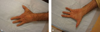
Figure 1: Intraoperative image demonstrating direct reduction and temporary K-wire fixation of an intra-articular phalangeal fracture. Note the meticulous manipulation of fragments to restore articular congruity prior to definitive plating.
Fixation
The choice of fixation method depends on fracture pattern, stability, bone quality, and surgeon preference. The goal is stable fixation that permits early mobilization.
-
K-wires:
- Temporary: For holding reduction while definitive fixation is applied.
- Definitive: For stable, non-comminuted fractures (e.g., transverse phalangeal fractures, some oblique fractures) or small avulsion fractures. Cross-pinning or parallel pinning techniques are common. Smooth K-wires are preferred for joints to allow motion, while threaded K-wires provide more grip in bone but can be difficult to remove and potentially damage soft tissues.
- K-wires are less suitable for articular reconstruction alone due to limited compressive force.
-
Mini-Screws and Plates:
- Lag Screws: Used for interfragmentary compression in long oblique or spiral fractures. Provides absolute stability, allowing primary bone healing. Requires adequate fragment size.
- Neutralization Plates: Protect lag screw constructs from bending, rotation, and shear forces.
- Reconstruction/Contour Plates: Used for comminuted fractures or articular surface reconstruction where anatomical contouring is essential.
- Locking Plates: Offer improved stability, especially in comminuted fractures or osteoporotic bone, by providing angular stability between the screw and plate. Useful when absolute stability cannot be achieved with traditional lag screws.
- Tension Band Wiring: Effective for avulsion fractures where a tendon or ligament pulls a bone fragment (e.g., mallet finger fracture, profundus avulsion). The wire converts tensile forces into compressive forces at the fracture site.
-
External Fixation:
- Indicated for highly comminuted, open fractures with significant soft tissue loss, unstable dislocations, or as a temporary measure in polytrauma.
- Can provide stability while allowing access to soft tissues for dressing changes. However, frame placement can interfere with early motion.
Closure
After achieving satisfactory fixation (confirmed with fluoroscopy and clinical assessment of stability and ROM if appropriate), the wound is irrigated.
*
Soft Tissue Repair:
Any incised retinacular structures, collateral ligaments, or tendon sheaths should be meticulously repaired. This is critical for preventing adhesions and restoring normal mechanics.
*
Layered Closure:
Subcutaneous tissues are approximated, followed by skin closure with fine, non-absorbable sutures to minimize scar formation.
*
Dressing:
A sterile, bulky soft dressing is applied, often incorporating a volar or dorsal splint to protect the repair while allowing for controlled motion based on the post-operative protocol. The "safe position" (MP flexion, IP extension) is often used for immobilization to prevent collateral ligament shortening at the MP joint.
Complications & Management
Despite meticulous surgical technique and aggressive rehabilitation, complications can arise, significantly challenging the achievement of optimal TAM. Understanding their incidence and implementing effective salvage strategies is crucial.
| Complication | Incidence | Salvage Strategies |
|---|---|---|
| Stiffness/Contracture | Very common (20-70%, depending on injury) | Aggressive hand therapy, dynamic/static progressive splinting, serial casting, steroid injections (peritendinous), tenolysis (for tendon adhesions), capsulotomy/arthrolysis (for joint contracture), staged reconstructive procedures. Prevention with early motion is key. |
| Nonunion/Malunion | 5-15% (higher in comminuted/open fractures) | Nonunion: Revision ORIF with bone grafting (autograft/allograft), electrical stimulation. Malunion: Corrective osteotomy (esp. for rotational/angular deformity), joint arthrodesis (if joint arthritis severe). |
| Infection | 1-5% (higher in open fractures) | Debridement, irrigation, antibiotics (IV then oral), hardware removal if unstable or infected. For osteomyelitis, aggressive debridement and prolonged antibiotics. |
| Hardware-Related Issues | 5-20% (prominence, migration, breakage) | Hardware removal (after healing), revision fixation if instability persists. Prominent hardware may interfere with tendon gliding or cause pain, requiring removal once bone healing is sufficient. |
| Complex Regional Pain Syndrome (CRPS) | 2-5% (variable reporting) | Multidisciplinary approach: pain management (medications, nerve blocks), aggressive physical/occupational therapy (desensitization, graded motor imagery), psychological support. Early diagnosis and treatment are critical for prognosis. |
| Tendon Adhesions | Very common post-trauma/surgery | Aggressive early active/passive ROM, scar massage, dynamic splinting, tenolysis (surgical release of adhesions). |
| Post-Traumatic Arthritis | 10-30% (higher in intra-articular fractures) | Joint fusion (arthrodesis) for pain relief and stability (loss of motion), joint replacement (arthroplasty, e.g., for MP joints), resection arthroplasty (e.g., for some PIP joints). Conservative management (NSAIDs, injections) initially. |
| Neurovascular Injury | <1% (iatrogenic) to 10-20% (original trauma) | Acute: Microsurgical repair (nerve/artery). Chronic nerve injury: Neurolysis, nerve grafting, tendon transfers for functional deficits. Vascular: Thrombectomy, bypass, or observation for vasospasm. |
| Scar Sensitivity/Contracture | Common | Scar massage, silicone sheeting, desensitization techniques, corticosteroid injections for hypertrophic scars. Z-plasty or other reconstructive plastic surgery for severe contractures. |
Detailed Management Strategies:
-
Stiffness and Contracture:
- Prevention is Paramount: This begins with atraumatic surgical technique, stable fixation allowing early motion, and immediate, specialized hand therapy.
- Early Intervention: Once stiffness is noted, escalation of therapy, custom splinting (dynamic or static progressive), and serial casting are indicated.
-
Surgical Salvage:
- Tenolysis: Surgical release of adhesions around tendons. Must be followed by extremely aggressive and immediate rehabilitation.
- Capsulotomy/Arthrolysis: Surgical release of a contracted joint capsule or lysis of intra-articular adhesions. Often performed concurrently with tenolysis.
- Staged Reconstruction: In severe cases, multiple procedures may be required.
-
Nonunion and Malunion:
- Nonunion: Failure of bone healing after an adequate period. Requires revision surgery, often with debridement of the nonunion site, stable internal fixation, and bone grafting (autogenous cancellous bone graft remains the gold standard).
-
Malunion:
Healing in an unacceptable position (angulation, rotation, shortening).
- Rotational Malunion: Most disabling, leading to finger scissoring. Requires corrective osteotomy and rigid internal fixation.
- Angular Malunion: Can alter tendon mechanics and joint loading. Corrective osteotomy.
- Articular Malunion: Leads to post-traumatic arthritis. Early stages managed conservatively; severe cases may require arthrodesis or arthroplasty.
-
Infection:
- Managed based on severity. Superficial infections may respond to oral antibiotics. Deep infections (osteomyelitis) require aggressive surgical debridement, tissue sampling for culture, intravenous antibiotics tailored to sensitivities, and potentially hardware removal if it acts as a nidus for infection.
-
Hardware-Related Issues:
- Prominent hardware can irritate soft tissues, cause pain, or impede tendon gliding. If bone healing is secure, symptomatic hardware should be removed. In some cases, hardware breakage may require revision fixation.
-
Complex Regional Pain Syndrome (CRPS):
- A severe, debilitating neuroinflammatory condition characterized by pain, swelling, allodynia, sudomotor changes, and trophic changes.
- Management: Multidisciplinary approach involving pain specialists (nerve blocks, neuromodulation, medications like gabapentinoids, NSAIDs, antidepressants), hand therapists (desensitization, graded motor imagery, pain education), and psychological support. Early diagnosis and aggressive treatment are paramount.
-
Post-Traumatic Arthritis:
- A common long-term sequela, particularly after intra-articular fractures or malunion.
- Management: Initial conservative management (NSAIDs, activity modification, steroid injections). For end-stage painful arthritis, surgical options include arthrodesis (joint fusion, providing pain relief and stability but sacrificing motion) or arthroplasty (joint replacement, preserving some motion but with potential long-term issues).
Post-Operative Rehabilitation Protocols
The success of hand injury treatment, particularly in optimizing TAM, hinges critically on a well-structured, individualized, and diligently followed post-operative rehabilitation protocol. This process demands close collaboration between the surgeon and a specialized hand therapist. The protocol must be tailored to the specific injury, the stability of the surgical fixation, and patient compliance.
Phase 1: Immediate Post-Operative (Day 0-2 weeks)
Goals:
* Control pain and edema.
* Protect surgical repair/fixation.
* Initiate earliest possible controlled motion.
* Educate patient on precautions and home exercises.
Interventions:
*
Edema Control:
Immediate elevation of the hand above the heart (e.g., slings, pillows). Gentle compression bandages or edema gloves.
*
Pain Management:
Pharmacological intervention as prescribed. Regional anesthesia may provide prolonged analgesia.
*
Wound Care:
Maintain clean, dry dressings. Monitor for signs of infection. Suture removal typically at 10-14 days.
*
Immobilization:
Custom-fabricated splint (thermoplastic) or cast to protect the repair while allowing for controlled motion of specific joints. The "safe position" (MP flexion 70-90 degrees, IP extension) is often used for MP joint protection to prevent collateral ligament shortening.
*
Controlled Active Motion (AROM):
* For stable fixation (e.g., rigidly fixed intra-articular fractures): Gentle AROM exercises for the involved and adjacent joints, ensuring no excessive stress on the repair. This may involve specific blocking exercises (e.g., MP blocking to isolate IP flexion).
* For tendon repairs: Specific early protected motion protocols (e.g., Kleinert, Duran, Washington, or various active motion protocols) under the guidance of a hand therapist, typically involving dynamic splinting.
* For unstable fixation or soft tissue reconstruction, passive ROM may be delayed, or only AROM to uninvolved joints allowed.
*
Patient Education:
Thorough instruction on home exercise program, activity modifications, signs of complications, and importance of adherence.
Phase 2: Early Mobilization (2-6 weeks post-op)
Goals:
* Gradually increase AROM and introduce PROM.
* Prevent adhesions and stiffness.
* Manage scar tissue.
* Maintain strength of uninvolved muscles.
Interventions:
*
Progressive ROM:
Gradually increase the intensity and range of active and passive exercises.
*
Active Range of Motion (AROM):
Continue with blocking exercises, full fist, hook fist, flat fist, composite flexion/extension.
*
Passive Range of Motion (PROM):
Gentle, pain-free passive stretches, performed by the therapist or guided by the patient. Overpressure may be applied cautiously.
*
Dynamic and Static Progressive Splinting:
For persistent stiffness or developing contractures, specialized splints (e.g., outrigger splints for extension, flexion slings) provide a constant, low-load stretch to gain motion.
*
Scar Management:
Scar massage, silicone sheeting, and elastomer inserts to desensitize the scar, improve elasticity, and prevent hypertrophy.
*
Edema Management:
Continue elevation, compression garments.
*
Intrinsic/Extrinsic Gliding:
Specific exercises to promote differential gliding of flexor and extensor tendons.
*
Light Gripping/Pinching:
May be introduced for very stable fixation towards the end of this phase, with caution.
Phase 3: Intermediate Phase (6-12 weeks post-op)
Goals:
* Restore full, pain-free ROM.
* Initiate progressive strengthening.
* Improve grip and pinch strength.
* Enhance fine motor coordination.
Interventions:
*
Full ROM:
Work towards achieving full AROM and PROM for all involved joints.
*
Strengthening:
*
Light Resistance:
Begin with light resistive exercises using therapeutic putty, rubber bands, or light weights.
*
Grip and Pinch:
Progress from gentle to more challenging grip and pinch strengthening exercises.
*
Fine Motor Coordination:
Introduce activities requiring dexterity, such as manipulating small objects, pegboards, or writing.
*
Functional Activities:
Incorporate tasks that mimic ADLs and work-related activities.
*
Proprioception:
Exercises to improve joint position sense.
*
Weaning from Splints:
Gradually decrease reliance on protective splints, using them primarily for strenuous activities or at night.
Phase 4: Advanced Phase and Return to Activity (>12 weeks post-op)
Goals:
* Maximize strength, endurance, and functional capacity.
* Return to work, sports, and desired activities.
* Long-term monitoring and problem-solving.
Interventions:
*
Progressive Resistance:
Continue to advance strengthening exercises to match pre-injury demands.
*
Endurance Training:
Activities that sustain muscular effort over time.
*
Sport-Specific/Work Hardening:
Tailored rehabilitation programs designed to meet the demands of the patient's specific sport or occupation.
*
Patient Education:
Ongoing education regarding injury prevention, proper body mechanics, and self-management strategies for any residual symptoms.
*
Outcome Measurement:
Objective assessment of TAM, grip strength, pinch strength, and patient-reported outcome measures (PROMs) like the QuickDASH or MHQ to quantify functional recovery.
Key Principles of Rehabilitation:
- Individualization: Protocols must be customized to the specific patient and injury.
- Early, Controlled Motion: The single most important factor for preventing stiffness and achieving TAM.
- Communication: Constant dialogue between the surgeon, hand therapist, and patient.
- Patient Adherence: Crucial for success; requires clear instructions and motivation.
- Gradual Progression: Exercises and activities should be advanced systematically, respecting tissue healing.
- Pain and Edema Management: Aggressive control of these factors to facilitate participation in therapy.
Summary of Key Literature / Guidelines
The pursuit of optimal TAM after hand injury is deeply rooted in established principles and continuously refined by evolving research. Several foundational concepts and guidelines inform modern surgical and rehabilitative practices.
Foundational Principles
-
AO Principles: The Arbeitsgemeinschaft für Osteosynthesefragen (AO Foundation) principles of fracture management are paramount:
- Anatomical Reduction: Especially crucial for articular surfaces to prevent post-traumatic arthritis.
- Stable Internal Fixation: Provides biomechanical stability allowing for early, controlled motion.
- Preservation of Blood Supply: Minimizing soft tissue stripping during surgical exposure.
-
Early Active Mobilization:
The cornerstone for preventing stiffness and optimizing TAM.
These principles, while originally developed for long bones, have been rigorously applied and adapted to the unique challenges of the hand.
-
Importance of Early Mobilization: Extensive literature supports that early, protected active motion after stable fixation significantly reduces the incidence and severity of post-operative stiffness and tendon adhesions. Studies by authors such as Duran and Houser, and Kleinert for flexor tendon repairs, revolutionized rehabilitation by demonstrating improved outcomes with early active and passive motion protocols compared to prolonged immobilization.
Specific Injury Guidelines
-
Intra-articular Fractures:
- Phalangeal Pilon Fractures (P2 Pilon): These are particularly challenging. Studies emphasize the need for anatomical reduction of the articular surface (often requiring open reduction and small fragment fixation with mini-plates/screws or K-wires) and stable fixation to allow early PIP joint motion. Long-term results often show some degree of residual stiffness despite optimal management, highlighting the difficulty in achieving full TAM.
- Metacarpal Head Fractures: Anatomical reduction and rigid fixation are crucial to preserve joint congruity and sagittal band integrity. Early motion protocols are similar to phalangeal injuries.
-
Tendon Repairs:
- Flexor Tendon Repairs (Zone II - "No Man's Land"): Traditionally associated with high rates of adhesions. Modern repairs utilize multi-strand core sutures and epitendinous sutures to achieve sufficient strength for early active motion protocols (e.g., early active place-and-hold, controlled active motion protocols). Outcomes are directly correlated with adherence to strict rehabilitation protocols.
- Extensor Tendon Repairs: Management varies by zone. Immobilization in extension is common for extensor lag, but early active motion is increasingly advocated for specific zones with stable repairs to prevent adhesions and maintain gliding.
-
Ligamentous Injuries:
- Ulnar Collateral Ligament (UCL) of the Thumb MP Joint: Complete ruptures (Stener lesion) require surgical repair. Early protected motion, often in a custom splint, is crucial.
- PIP Collateral Ligament Injuries: Stable sprains are managed non-operatively with buddy taping and early motion. Unstable tears or avulsions may require surgical repair followed by protected mobilization.
Role of Hand Therapy
The literature consistently highlights the indispensable role of certified hand therapists (CHTs) in achieving optimal outcomes. A specialized hand therapist is trained to:
* Assess functional deficits precisely.
* Fabricate custom splints (static, dynamic, static progressive) to protect repairs and augment motion.
* Implement advanced rehabilitation techniques (scar massage, desensitization, specific therapeutic exercises).
* Provide continuous feedback to the surgeon regarding patient progress and potential complications.
Current Trends & Future Directions
- Minimally Invasive Techniques: While challenging in the hand due to small structures, advancements in arthroscopy and percutaneous fixation are improving visualization and reducing soft tissue dissection.
- Advanced Imaging: High-resolution CT and MRI continue to refine pre-operative planning.
- Biologics: The role of biologics (e.g., PRP, stem cells) in enhancing bone and soft tissue healing in acute hand trauma is under investigation but not yet standard practice.
- Patient-Reported Outcome Measures (PROMs): Increasingly used to objectively quantify functional recovery from the patient's perspective (e.g., QuickDASH, Michigan Hand Outcomes Questionnaire - MHQ).
- Prevention of CRPS: Ongoing research into early diagnosis and aggressive multi-modal treatment strategies.
- Digital Health Technologies: Wearable sensors and telerehabilitation are emerging tools to monitor patient progress and provide remote guidance, potentially enhancing adherence and access to specialized care.
In summary, achieving optimal TAM after hand injury remains a significant challenge. Success demands a comprehensive strategy encompassing accurate diagnosis, judicious selection of surgical or non-operative management, precise surgical execution respecting the delicate anatomy, stable fixation, and, critically, an individualized, aggressive, and early post-operative rehabilitation program involving a dedicated hand therapist. Adherence to established principles and an awareness of evolving literature are essential for maximizing patient function.
