
Introduction & Epidemiology
Extraarticular two-part surgical neck fractures of the proximal humerus represent a significant portion of upper extremity trauma, particularly in older adults. These fractures are characterized by a transverse or oblique fracture line distal to the humeral head articular surface but proximal to the shaft, with no extension into the greater or lesser tuberosities. The designation "2-part" indicates separation between the humeral head/tuberosity block and the humeral shaft, with the tuberosities remaining attached to the humeral head. "Impaction" refers to a stable variant where the metaphyseal bone of the humeral head is driven into the cancellous bone of the shaft, creating intrinsic stability.
The incidence of proximal humerus fractures is approximately 70-80 per 100,000 person-years, with a marked increase in prevalence in the elderly population, especially postmenopausal women, often associated with osteoporosis and low-energy falls. While multi-part fractures typically raise concerns for avascular necrosis (AVN) and complex reconstruction, two-part surgical neck fractures, particularly those that are impacted, generally carry a better prognosis regarding union and functional outcomes. However, malunion can still lead to significant functional impairment, pain, and reduced range of motion. The management spectrum ranges from non-operative treatment with early mobilization to operative fixation, with the decision-making process largely dictated by fracture stability, displacement, angulation, and patient-specific factors such as age, comorbidities, and functional demands. This review focuses on the operative management of impacted surgical neck fractures, emphasizing the principles of reduction and stable impaction fixation, primarily through lag screw constructs.
Surgical Anatomy & Biomechanics
A thorough understanding of the regional anatomy and biomechanics is paramount for successful surgical management of proximal humeral fractures.
Bony Anatomy
The
proximal humerus
comprises the humeral head, anatomical neck, surgical neck, greater tuberosity, lesser tuberosity, and bicipital groove.
* The
humeral head
is articular, covered by hyaline cartilage, and articulates with the glenoid.
* The
anatomical neck
lies just distal to the articular margin of the humeral head.
* The
surgical neck
is the common site for fractures, located distal to the tuberosities and anatomical neck, at the junction of the wider metaphyseal bone and the narrower diaphysis.
* The
greater tuberosity
is the attachment site for the supraspinatus, infraspinatus, and teres minor tendons.
* The
lesser tuberosity
is the attachment site for the subscapularis tendon.
Soft Tissue & Neurovascular Anatomy
- Deltoid Muscle: The deltoid covers the shoulder joint superiorly, anteriorly, and posteriorly. Its innervation is by the axillary nerve. Different surgical approaches split or detach portions of the deltoid.
- Rotator Cuff: Comprises the supraspinatus, infraspinatus, teres minor, and subscapularis. These muscles are critical for shoulder stability and motion. Their integrity and potential injury during fracture or surgery influence rehabilitation.
- Axillary Nerve: This nerve (C5-C6) arises from the posterior cord of the brachial plexus. It courses inferiorly and posteriorly, exiting the quadrangular space (bounded by the teres minor superiorly, teres major inferiorly, long head of triceps medially, and surgical neck of the humerus laterally). It then wraps around the surgical neck of the humerus, approximately 5-7 cm distal to the lateral acromial edge, supplying the deltoid and teres minor muscles and providing sensory innervation to the lateral shoulder. Protection of the axillary nerve is critical during any surgical approach to the proximal humerus, especially during screw insertion or retraction.
- Radial Nerve: Although generally more distal, the radial nerve is a concern with more distal extension of surgical neck fractures or during extensive exposure.
- Musculocutaneous Nerve: Located more medially, it innervates the biceps and brachialis muscles.
- Arterial Supply: The humeral head receives its primary blood supply from the ascending branch of the anterior humeral circumflex artery (arcuate artery) and branches of the posterior humeral circumflex artery. The integrity of this blood supply is crucial to prevent AVN, although in two-part surgical neck fractures, the blood supply to the head is often preserved due to intact tuberosity attachments.
Biomechanics of Impaction Fixation
In an impacted surgical neck fracture, the metaphyseal cancellous bone of the humeral head is driven into the cancellous bone of the humeral shaft. This creates inherent stability through bone-on-bone compression. The goal of fixation, particularly with lag screws, is to augment this intrinsic stability, maintain reduction, and enhance interfragmentary compression. Lag screws are ideal for achieving this as they compress the two fragments across the fracture line. The oblique nature of the fracture plane in many impacted fractures allows for a longer screw trajectory and thus greater purchase and compression. The forces acting on the proximal humerus (deltoid pull, rotator cuff action) must be counteracted by a stable construct to allow early mobilization and prevent loss of reduction.
Indications & Contraindications
The decision to proceed with operative fixation for an impacted two-part surgical neck fracture is nuanced, weighing patient factors, fracture characteristics, and the potential for functional compromise with non-operative management.
General Considerations
Impaction inherently confers stability, and many such fractures can be successfully managed non-operatively. The primary indications for operative intervention revolve around unacceptable displacement or angulation that is likely to lead to poor functional outcomes or a high risk of secondary displacement.
- Intrinsically Stable Fractures: As noted, impacted fractures are intrinsically stable due to metaphyseal impaction. They typically respond well to gentle early mobilization.
- Deformity Tolerance: The shoulder has a remarkable ability to compensate for mild-moderate angulation (up to 30-45 degrees varus, 20 degrees valgus, 20-30 degrees apex anterior/posterior). Significant deformity, however, may lead to impingement, pain, and restricted range of motion.
- Patient Factors: Age, activity level, functional demands, hand dominance, and comorbidities all influence the treatment decision. A young, active patient with significant angulation may warrant fixation, whereas an elderly, sedentary patient might be better served by non-operative care even with similar angulation.
- Risk of Delayed Displacement: Although impacted fractures are stable, certain patterns (e.g., highly comminuted surgical neck, reverse obliquity) can be at higher risk of secondary displacement, even after initial impaction.
Operative Indications
-
Displacement:
- Greater than 1 cm displacement.
- Significant rotational malalignment (e.g., >45 degrees).
-
Angulation:
- Greater than 45 degrees of angulation (varus or valgus).
- Apex anterior or posterior angulation exceeding 30 degrees, particularly in younger, active patients.
- Significant valgus impaction that creates an abduction deformity and impingement.
- Non-reducible Deformity: Inability to achieve acceptable alignment with closed reduction maneuvers.
- Patient Demands: Younger, high-demand patients where even mild residual deformity is unacceptable.
- Open Fractures: Although rare for this fracture type, any open fracture necessitates operative debridement and fixation.
- Vascular Injury: Associated neurovascular injury requiring exploration and repair (though fixation usually follows primary repair).
Relative Contraindications
- Non-displaced or Minimally Displaced Fractures: If angulation is within acceptable limits and the fracture is stable to stress, non-operative management is typically preferred.
- Elderly, Low-Demand Patients: With significant comorbidities or limited life expectancy, even some displaced fractures may be better managed non-operatively to avoid surgical risks.
- Severe Osteoporosis: Poor bone quality may preclude stable screw fixation, leading to implant pull-out. In such cases, alternative fixation (e.g., plate osteosynthesis, arthroplasty) or non-operative treatment might be considered, though specific impaction screws can sometimes achieve surprising stability.
- Active Infection: Absolute contraindication to elective internal fixation.
- Poor Soft Tissue Envelope: Compromised soft tissues or significant skin conditions that increase infection risk.
Summary Table: Operative vs. Non-Operative Indications
| Feature | Non-Operative Treatment (Preferred) | Operative Treatment (Considered) |
|---|---|---|
| Displacement | Minimally displaced (< 1 cm) | > 1 cm displacement |
| Angulation | Valgus < 20°, Varus < 45°, Apex Ant/Post < 30° | Valgus > 20°, Varus > 45°, Apex Ant/Post > 30° |
| Rotational Malalignment | < 45° | > 45° or causing impingement |
| Fracture Type | Stable, well-impacted surgical neck fracture, particularly varus | Unstable fracture pattern or risk of secondary displacement |
| Patient Age/Demand | Elderly, sedentary, low-demand, significant comorbidities | Young, active, high-demand, few comorbidities |
| Bone Quality | Good to moderate, allowing for inherent stability | Good, allowing for stable screw fixation (if screws chosen) |
| Associated Injuries | Isolated fracture | Open fracture, neurovascular compromise, polytrauma requiring early mobilization |
| Ability to Reduce Closed | Successful closed reduction to acceptable alignment | Unsuccessful closed reduction or unstable after reduction |
Pre-Operative Planning & Patient Positioning
Meticulous pre-operative planning is crucial for predictable outcomes in extraarticular surgical neck impaction fixation.
Pre-Operative Assessment
- Clinical Evaluation: Assess neurovascular status (axillary nerve function, radial pulse, capillary refill). Document pre-injury functional status and comorbidities.
-
Imaging:
- Standard Radiographs: True AP, scapular Y, and axillary views are essential. These provide initial assessment of fracture pattern, displacement, and angulation. Specific attention should be paid to the degree and type of impaction.
- CT Scan: Highly recommended, especially for difficult-to-visualize fractures or when rotational displacement is suspected. A CT scan with 3D reconstructions can precisely delineate the fracture morphology, degree of comminution, and bone quality, aiding in implant selection and surgical strategy. It helps confirm if the fracture truly is a "2-part, impacted surgical neck" as opposed to a 3-part or 4-part fracture with occult tuberosity involvement.
- MRI: Rarely indicated acutely but may be useful if there is concern for significant associated soft tissue injury (e.g., rotator cuff tear) that might alter rehabilitation.
Templating and Implant Selection
- Implant Choice: For impacted two-part surgical neck fractures where the primary goal is to augment impaction and provide compression, headless or partially threaded cancellous lag screws are the implants of choice. The choice of diameter (e.g., 4.0 mm, 4.5 mm, 6.5 mm) depends on bone quality and the size of the humeral head.
- Screw Trajectory: Templating helps determine the optimal screw trajectory to maximize purchase in both the humeral head fragment and the shaft fragment while avoiding articular penetration. Aim for screws to cross the fracture perpendicularly to the fracture plane for maximum compression.
- Screw Length: Accurate measurement from radiographs or CT scans helps estimate appropriate screw lengths to avoid articular penetration proximally and unnecessary protrusion distally. Fluoroscopy during surgery will confirm final length.
- Alternative Implants: In cases where screw fixation alone is deemed insufficient due to a less stable impaction, or if there's a concern for propagation, a supplementary locking plate (e.g., PHILOS plate) can be considered, especially in osteoporotic bone. However, for true impaction fixation, the focus remains on lag screws.
Patient Positioning and Surgical Preparation
- Anesthesia: General anesthesia is typically employed. A regional nerve block (interscalene brachial plexus block) can be highly beneficial for post-operative pain management.
-
Patient Position:
The patient is usually positioned in a
beach chair position
(semisitting), with the torso elevated 30-45 degrees.
- The head is secured and slightly turned away from the operative side.
- The involved arm is draped free to allow for full range of motion, particularly adduction, extension, and internal/external rotation, which are critical for reduction maneuvers.
- Careful padding of all pressure points (sacrum, heels, opposite elbow) is essential to prevent nerve palsies or skin breakdown.
- Fluoroscopy access is critical. The C-arm should be positioned to allow unobstructed views in AP and axillary planes without repositioning the patient or the C-arm during the procedure. This typically means starting with the C-arm at the foot of the bed for AP views and swinging it in for axillary views, or having it positioned laterally.
- Surgical Preparation: The entire shoulder, upper arm, and axilla are prepped and draped in a sterile fashion. A sterile tourniquet is generally not used for shoulder surgery due to the risk of nerve injury and muscle ischemia, especially given the typically short operative time for this procedure. Infiltration with local anesthetic (e.g., bupivacaine with epinephrine) can aid hemostasis and provide further post-operative analgesia.
Detailed Surgical Approach / Technique
The goal of surgical fixation for impacted two-part surgical neck fractures is to achieve anatomical reduction, maintain impaction, and provide stable compression using lag screws while minimizing soft tissue disruption and neurovascular injury.
Surgical Approach Selection
Two primary approaches are commonly used:
1.
Deltopectoral Approach:
This is the workhorse approach for most proximal humerus fractures.
*
Advantages:
Allows excellent visualization of the anterior and medial aspects of the proximal humerus, protects the axillary nerve, and is extensile for potential plate fixation if needed. It follows an internervous plane between the deltoid (axillary nerve) and pectoralis major (medial and lateral pectoral nerves).
*
Disadvantages:
Limited access to the posterior and lateral aspects of the humeral head, especially for posteromedial screw trajectories.
2.
Deltoid-Splitting (Minimally Invasive Lateral) Approach:
*
Advantages:
Less soft tissue dissection, direct access to the lateral aspect of the humerus, good for direct screw placement from lateral.
*
Disadvantages:
Risk of axillary nerve injury if the split extends too far distally (limit to 5 cm from the acromion). Restricted visualization compared to the deltopectoral approach. More suitable for very minimally displaced or perfectly reduced fractures requiring direct lateral screw insertion.
For impaction fixation requiring precise reduction and compression, especially if multiple screws or specific trajectories are planned, the deltopectoral approach is generally favored for its superior control and safety.
Step-by-Step Surgical Technique (Deltopectoral Approach)
-
Incision:
- A longitudinal incision is made from the tip of the coracoid process extending distally for approximately 6-8 cm along the deltopectoral groove.
- Carefully incise the skin and subcutaneous tissue. Identify the cephalic vein, which lies within the deltopectoral groove. It is typically retracted medially with the pectoralis major.
- Internervous Plane: The deltopectoral interval is identified between the deltoid muscle (lateral) and the pectoralis major muscle (medial).
- Blunt dissection proceeds along this interval.
-
Exposure:
- Retract the deltoid laterally and the pectoralis major medially.
- Identify the clavipectoral fascia beneath the deltoid and pectoralis major. Incise it longitudinally.
- This exposes the underlying coracobrachialis and short head of biceps (medially) and the subscapularis tendon (laterally). The subscapularis is typically left intact for two-part surgical neck fractures.
- The anterior humeral circumflex vessels may be encountered and should be ligated or carefully cauterized.
-
Axillary Nerve Protection:
- Crucial step: The axillary nerve, often accompanied by the posterior humeral circumflex artery, courses around the surgical neck approximately 5-7 cm distal to the acromial edge.
-
-
Protect the nerve by:
- Limiting distal dissection.
- Using blunt dissection and careful retractors (e.g., blunt Hohmann retractors) placed directly on bone.
- Avoiding aggressive inferior retraction of the deltoid.
- Palpating the nerve and its associated vessels if direct visualization is difficult.
-
Fracture Reduction:
-
Disimpaction:
This is the key to successful reduction. The impacted fragments must first be gently disimpacted.
- Use a blunt elevator (e.g., periosteal elevator, small osteotome) or a K-wire to carefully lever the humeral head fragment off the shaft fragment. Avoid aggressive force to prevent comminution or iatrogenic fracture.
- Often, traction and gentle manipulation of the arm, coupled with disimpaction, are sufficient.
-
Reduction Maneuvers:
- The primary deforming forces are often the pectoralis major (pulling the shaft medially and anteriorly) and the rotator cuff (pulling the head into flexion and external rotation).
- Apply longitudinal traction to the arm.
- Use internal rotation to counteract the external rotation of the head (if present).
- Use an instrument (e.g., blunt hook, bone clamp, K-wire joystick) to manipulate the humeral head fragment.
- Manipulate the shaft fragment (often easier) by applying varus/valgus or anterior/posterior pressure.
- Proper reduction: After reduction, alignment should be correct in both sagittal and coronal planes. Rotational alignment must also be correct.
- Confirm reduction with fluoroscopy in AP and axillary views. Temporary K-wires can be used to hold the reduction if it is tenuous, especially important to prevent rotation.
-
Disimpaction:
This is the key to successful reduction. The impacted fragments must first be gently disimpacted.
-
Lag Screw Fixation:
- Principles: Impacted surgical neck fractures without comminution, if the fracture plane is oblique and long enough, may often be fixed satisfactorily with lag screws after closed reduction. Lag screws function by compressing the fragments together.
-
Drill Guide & K-wires:
Using a drill guide, insert one or two guide wires (e.g., 2.0 mm) from the lateral cortex of the humeral shaft, across the fracture line, and into the humeral head fragment.
- Trajectory: Aim for a trajectory that is as perpendicular as possible to the fracture plane to maximize compression. The oblique nature of surgical neck fractures often allows for this.
- Placement: Ensure wires are not too close to the articular surface (check fluoroscopically in both AP and axillary views). Aim for divergent screw trajectories to resist rotation.
-
Drilling:
- For fully threaded cancellous screws, overdrill the near cortex (shaft fragment) with a drill bit the same diameter as the screw.
- Underdrill the far cortex and humeral head fragment with a drill bit the diameter of the screw core.
- For partially threaded cancellous screws, the overdrilling of the near cortex is usually not necessary as the unthreaded portion acts as a gliding hole.
- Depth Gauge: Measure the length of the guide wire or drilled hole to determine the appropriate screw length.
- Tapping: If bone quality is dense, tap the far cortex and humeral head fragment to facilitate screw insertion and prevent stripping.
-
Screw Insertion:
Insert the chosen cancellous lag screws. As the screw is tightened, the threads in the humeral head engage, and the unthreaded portion in the shaft allows the head fragment to be drawn towards the shaft, creating compression across the fracture.
- Typically, two or three screws are sufficient, inserted in divergent paths to enhance rotational stability.
- Ensure adequate purchase in the humeral head.
- Final Fluoroscopy: Confirm screw position, length (no articular penetration), and maintenance of reduction in AP and axillary views.
-
-
- Stability Assessment: Gently stress the fracture under fluoroscopy to ensure robust fixation and maintenance of the reduction.
-
Wound Closure:
- Irrigate the wound thoroughly.
- Close the deep fascia (clavipectoral fascia) and subcutaneous layers.
- Close the skin with absorbable or non-absorbable sutures.
- Apply a sterile dressing. The arm is typically placed in a sling for comfort and initial protection.
Complications & Management
Despite meticulous surgical technique, complications can arise following fixation of two-part surgical neck impaction fractures. Prompt recognition and appropriate management are crucial for salvage.
Common Complications
| Complication | Incidence (Approximate) | Management / Salvage Strategies |
|---|---|---|
| Axillary Nerve Injury | 1-10% (transient), <1% (permanent) | Prevention is key: Limit distal dissection to >5 cm from acromion, careful retraction, blunt dissection. Management: Often transient neurapraxia. Observe for spontaneous recovery (up to 6-12 months). EMG/NCS studies if no recovery. Surgical exploration and neurolysis/repair for persistent deficits or iatrogenic transection. Bracing/sling for deltoid paralysis. |
| Loss of Reduction/Fixation Failure | 5-15% | Often due to inadequate initial reduction, poor bone quality, or premature aggressive rehabilitation. Management: Revision surgery with more robust fixation (e.g., locking plate), bone grafting for nonunion. In severe cases or poor bone stock, arthroplasty (hemiarthroplasty or reverse total shoulder arthroplasty) may be considered, particularly in older patients. |
| Nonunion/Malunion | 5-10% | Nonunion: Persistent pain and failure to unite after 6-9 months. Malunion: United in an unacceptable position (e.g., symptomatic impingement). Management: For symptomatic nonunion, revision fixation with bone grafting, potentially plate osteosynthesis. For malunion, corrective osteotomy or arthroplasty (hemiarthroplasty or reverse TSA) depending on age, deformity, and functional demands. |
| Avascular Necrosis (AVN) of Humeral Head | <5% (2-part fractures) | Incidence is much lower than in 3- or 4-part fractures as blood supply is often preserved. Management: Early stages: conservative management, pain control. Later stages: core decompression (rarely), hemiarthroplasty, or total shoulder arthroplasty depending on patient age, activity level, and glenoid status. |
| Infection (Superficial/Deep) | <2% (superficial), <1% (deep) | Prevention: Meticulous sterile technique, prophylactic antibiotics. Management: Superficial: oral antibiotics, wound care. Deep: surgical debridement, irrigation, cultures, IV antibiotics (based on sensitivities), potential implant removal if chronic and unstable. |
| Implant-Related Issues | 5-10% | Screw protrusion/articular penetration: Pain, chondral damage. Management: Early removal of offending screw(s). If significant damage, potential arthroscopic debridement or salvage with arthroplasty in chronic cases. Screw backout: Loss of fixation. Management: Revision fixation, often with plate osteosynthesis. |
| Stiffness/Adhesive Capsulitis | 10-20% | Common, particularly after prolonged immobilization. Prevention: Early, controlled range of motion. Management: Aggressive physical therapy, oral NSAIDs, intraarticular corticosteroid injections. If refractory, manipulation under anesthesia (MUA) or arthroscopic capsular release. |
| Shoulder Impingement | 5-10% | Often secondary to malunion (e.g., excessive varus or anterior angulation). Management: Conservative initially (PT, NSAIDs, injections). If persistent, arthroscopic subacromial decompression (acromioplasty) and potential revision of malunion or arthroplasty. |
| Complex Regional Pain Syndrome (CRPS) | Rare (<1%) | Prevention: Adequate pain control, early mobilization. Management: Multidisciplinary approach: physical therapy, pain management specialists, nerve blocks, medications (gabapentin, amitriptyline), psychological support. Early diagnosis and treatment are crucial. |
General Principles of Complication Management
- Early Recognition: Vigilant post-operative monitoring and prompt investigation of any new or worsening symptoms.
- Imaging: X-rays, CT scans, or MRI may be necessary to identify the cause of symptoms.
- Multidisciplinary Approach: Involve physical therapists, pain specialists, and other subspecialists as needed.
- Patient Communication: Clear and empathetic communication with the patient regarding expectations and potential outcomes of salvage procedures.
Post-Operative Rehabilitation Protocols
Post-operative rehabilitation is as critical as the surgery itself for optimizing functional outcomes and preventing complications such as stiffness or loss of reduction. The principles focus on protection of the repair, gradual restoration of motion, and progressive strengthening. Given the intrinsic stability of impacted fractures augmented by fixation, gentle early mobilization is key.
Phase 1: Immobilization and Early Passive Motion (Weeks 0-4/6)
- Goal: Protect the fracture, manage pain, prevent stiffness, and initiate early healing.
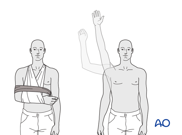
- Immobilization: The arm is typically placed in a sling for comfort and protection. The sling can be removed for hygiene and exercises.
-
Early Gentle Mobilization:
- Pendulum exercises: Started within the first few days, with the patient leaning forward, allowing the arm to hang freely and swing gently in small circles (clockwise and counter-clockwise) and anterior-posterior motions.
-
Passive Range of Motion (PROM):
Initiated with the assistance of a therapist or the contralateral hand.
- Forward flexion: To 90-120 degrees, respecting pain.
- External rotation: To 20-30 degrees (neutral to slight external rotation), being cautious not to overstress the healing bone, especially if the fracture involves posterior structures.
- Internal rotation: Performed by having the patient place their hand on their abdomen or lower back.
- Elbow, Wrist, Hand Exercises: Actively encouraged multiple times a day to prevent stiffness in these joints.
- Weight-Bearing Restrictions: Absolutely no weight-bearing, lifting, pushing, or pulling with the affected arm.
- Ice/Heat: Apply ice for pain and swelling, then heat prior to exercises to increase tissue extensibility.
Phase 2: Active-Assistive and Active Range of Motion (Weeks 4/6 - 12)
- Goal: Gradually increase active shoulder motion, restore neuromuscular control, and prepare for strengthening.
- Progression Criteria: Clinical and radiographic evidence of early fracture healing (callus formation), minimal pain.
- Active-Assistive Range of Motion (AAROM): Progress from PROM to AAROM using assistive devices (e.g., stick, pulley system) or the unaffected arm.
-
Active Range of Motion (AROM):
Once AAROM is comfortable and fracture stability is assured, begin AROM in all planes.
- Focus on achieving full functional range of motion, particularly forward flexion, abduction, and external/internal rotation.
- Avoid sudden movements or heavy lifting.
- Isometric Strengthening: Initiate gentle isometric exercises for the deltoid and rotator cuff muscles (subscapularis, supraspinatus, infraspinatus, teres minor) against resistance, avoiding painful contractions.
- Scapular Stabilization: Exercises to strengthen scapular retractors and depressors (rhomboids, middle/lower trapezius, serratus anterior) are incorporated.
Phase 3: Progressive Strengthening and Functional Return (Weeks 12 - 6 months+)
- Goal: Restore full strength, power, endurance, and return to functional activities.
- Progression Criteria: Full, pain-free active range of motion, satisfactory fracture healing on radiographs.
-
Resistive Strengthening:
- Begin with light resistance bands or light weights.
- Progress systematically, strengthening all major shoulder muscle groups (deltoid, rotator cuff, biceps, triceps, scapular stabilizers).
- Focus on eccentric and concentric exercises.
- Proprioception and Neuromuscular Control: Incorporate balance and coordination exercises (e.g., rhythmic stabilization, plyometrics for athletes).
- Sport-Specific/Work-Specific Training: For athletes or individuals with demanding occupations, integrate specific drills and simulations relevant to their activities.
- Return to Activity: Gradual return to sports or heavy labor, typically not before 4-6 months, and only after achieving near-normal strength and motion and complete radiographic healing. Contact sports or activities with high risk of re-injury may require longer.
Important Considerations for Rehabilitation
- Pain Management: Adequately manage pain throughout the rehabilitation process to allow for effective participation.
- Individualization: Protocols must be tailored to the individual patient's fracture stability, bone quality, age, comorbidities, and functional goals.
- Patient Compliance: Emphasize the importance of adherence to the rehabilitation program.
- Surgeon-Therapist Communication: Close collaboration between the surgeon and physical therapist is essential for safe and effective progression.
- Beware of Impingement: If malunion or significant callus forms, impingement can limit motion. This may require modifications to the protocol or further intervention.
Summary of Key Literature / Guidelines
The management of two-part surgical neck fractures has evolved with advances in understanding fracture biomechanics, implant technology, and rehabilitation. While the focus of this text is on surgical fixation for impacted variants, a broader understanding of the literature informing the overall treatment algorithm is beneficial.
Early series, primarily focused on non-operative management of impacted fractures, demonstrated acceptable outcomes with sling immobilization and early mobilization. Neer's classification (1970) provided a framework, highlighting the relative stability of two-part fractures. Studies by Court-Brown et al. (2001) and Gruson et al. (2008) have further elaborated on the epidemiology and outcomes, often showing good results for minimally displaced or impacted fractures managed non-operatively.
However, a subset of these fractures with significant displacement or angulation benefits from surgical intervention. The challenge has always been to achieve stable fixation in often osteoporotic bone.
*
Lag screw principles
are well-established in fracture fixation, emphasizing compression across the fracture site. For oblique surgical neck fractures, appropriately placed lag screws can convert bending forces into compression, enhancing stability. While specific large-scale randomized controlled trials (RCTs) exclusively comparing lag screw fixation for impacted 2-part surgical neck fractures versus non-operative management are limited due to the specific nature of this fracture type, the biomechanical principles are robust.
*
The AO Foundation
principles, fundamental to orthopedic trauma surgery, advocate for anatomical reduction and stable internal fixation allowing for early functional mobilization. For two-part surgical neck fractures, the AO classification 11-A2 (extraarticular, simple, metaphyseal) and 11-A3 (extraarticular, multi-fragmentary, metaphyseal) are relevant. Impacted simple metaphyseal fractures (akin to 11-A2) often lend themselves well to lag screw fixation, especially if the fracture plane is suitable.
*
Studies on minimally invasive techniques
for proximal humerus fractures, while often focusing on locking plates, reinforce the concept of preserving the biology of the fracture. Lag screw fixation, when performed through a limited approach, aligns with these principles.
*
Recent trends
in general proximal humerus fracture management suggest a preference for non-operative treatment for many stable, non-displaced fractures, particularly in the elderly. However, for those requiring intervention, the choice between various fixation methods (screws, plates, nails, arthroplasty) depends heavily on the fracture pattern, bone quality, and patient factors. For the specific niche of
impacted
two-part surgical neck fractures where the inherent stability is augmented, lag screw fixation stands as a biomechanically sound and often minimally invasive option, allowing for direct compression and early mobilization.
Guidelines from various trauma societies often emphasize:
1.
Careful patient selection:
Tailoring treatment to the individual.
2.
Anatomical reduction:
Crucial for preventing impingement and restoring function.
3.
Stable fixation:
To allow early motion and prevent loss of reduction.
4.
Early rehabilitation:
To avoid stiffness.
In conclusion, while the majority of impacted surgical neck fractures may be amenable to non-operative management, understanding the precise indications and executing a technically sound lag screw fixation procedure for appropriate candidates offers a reliable method for achieving anatomical reduction, stable union, and excellent functional recovery with careful post-operative rehabilitation.
