A 60-year-old female presents with a chief complaint of an intermittently painful mass on the medial border of the plantar medial arch in the left foot. It has been present for approximately 5 years but has not grown in size over the past 2 years. When pain occurs, it is only in shoes with an arch support. She is pain-free walking barefoot. Physical examination reveals a subcutaneous firm nodule along the medial cord of the plantar fascia. There are no overlying skin lesions. Tinel’s sign is negative. Plain x-rays (Figs. 5–31 and 5–32) and an MRI of the foot were obtained. Figures 5–33 and 5–34 are pregadolinium and Figure 5–35 is postgadolinium.
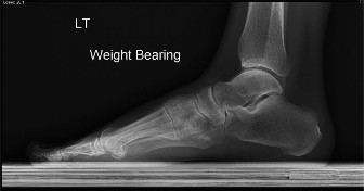
Figure 5–31 Lateral radiograph of the foot.
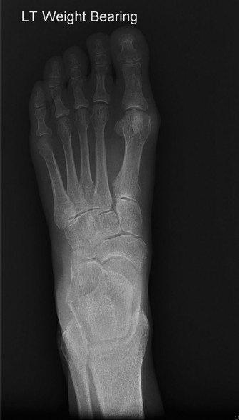
Figure 5–32 AP radiograph of the foot.
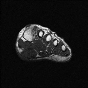
Figure 5–33 Coronal MRI pregadolinium contrast.
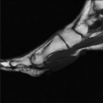
Figure 5–34 Sagittal MRI pregadolinium contrast.
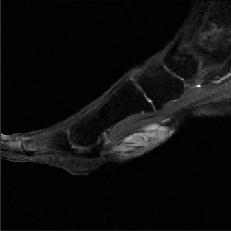
Figure 5–35 Sagittal MRI postgadolinium contrast.
The most appropriate treatment is:
- Referral to an orthopaedic oncologist for definitive management
- An excisional biopsy of the lesion
- To obtain a plain chest x-ray and plan for a wide margin resection of the lesion
- To obtain a needle biopsy of the lesion and await pathology findings for definitive management
- To consider a custom-molded orthotic to accommodate the lesion and provide reassurance
Discussion
The correct answer is (E). The patient has a plantar fibroma. Fibromas are typically solitary tumors located along the medial cord of the plantar fascia that contain dense, mature fibrocytes. In older patients, plantar fibromas may be associated with Dupuytren’s or Peyronie’s disease. Most often they are painless and require no treatment at all. When pain does occur, it is usually the result of the incongruous border rubbing against the insole of a shoe. Surgical excision is ill-advised in the vast majority of cases. Post surgery recurrence is very high because extensions of the lesion infiltrate the dermis and fascia making obtaining clean margins exceedingly difficult. Malignant degeneration of these lesions is essentially unheard of. An MRI can be obtained if the diagnosis is uncertain. Gadolinium will increase the specificity of the examination. A T1 image demonstrates low signal comparable
to muscle. The lesion will demonstrate variable enhancement with gadolinium. Soft tissue sarcomas of the foot and ankle do exist but are quite rare. The most common soft tissue sarcoma in the foot is a synovial cell sarcoma. These malignant tumors generally occur in patients younger than 30 years of age. There is a propensity for these tumors to metastasize to the lungs and wide margin surgical excision is required for treatment.
Objectives: Did you learn...?
Identify typical clinical presentation for a plantar fibroma? Treat a plantar fibroma?
