Clinodactyly is a condition that causes the fingers to curve towards the pinky finger. It is most common in the pinky finger and is often bilateral. The condition is usually harmless and does not require treatment.DR.MOHAMMAD HUTAIF,EMIAL
DEFINITION
Clinodactylyrefers to an abnormal about of radioulnar angulation of a digit ( >15 degrees). The small finger is most commonly observed.This condition is often bilateral.
ANATOMY
The finger consists of three phalanges (proximal, middle, and distal).The normal phalangeal physis is located at the proximal portion of each phalanx.
PATHOGENESIS
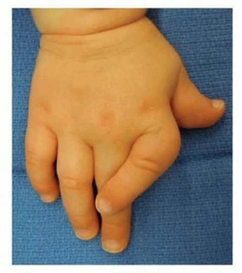
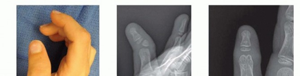
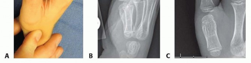
The angulation is result of abnormal development of one of the phalanges (most often the middle phalanx [p2]).Abnormal development of the phalanx may be due to an irregular physis (longitudinal bracket epiphysis). This may also be referred to as adelta phalanx.The tethering effect of the bracket epiphysis on the radial side of the finger causes abnormal growth of the phalanx resulting in a triangular or trapezoidal shape.Extra bones may be encountered.NATURAL HISTORYThe natural history of clinodactyly is variable and poorly documented, owing to the great number of cases that are asymptomatic and do not require treatment.Angulation may be stable or rapidly progressive at times of growth, depending on the extent of the involvement of the physis and/or presence of extra phalanges.PATIENT HISTORY AND PHYSICAL EXAM FINDINGSClinodactyly may be present at birth or develop during a period of growth (FIG 1). Clinodactyly is often bilateral in the small finger.Clinodactyly is an autosomal dominant condition with variable penetration. Involvement of the thumb is rare and is associated with varying syndromes.IMAGING AND OTHER DIAGNOSTIC STUDIESStandard radiographs (three views: anteroposterior[AP], lateral [LAT], and oblique [OBL]) of the hand and affected digit are sufficient to determine the area of involvement.Contralateral images are useful for comparison.Advanced imaging such as computed tomography (CT) is rarely needed. Magnetic resonance imaging (MRI)may be useful to delineate the shape of a bracket diaphysis.DIFFERENTIAL DIAGNOSISThe diagnosis of clinodactyly is straightforward; clinical examination and radiographs are sufficient to make the diagnosis.Associated syndrome should be screened for, these include Down, Rubinstein-Taybi, Apert, and Russell-Silver.NONOPERATIVE MANAGEMENTObservation may be considered for angulated digits that do not impair function. Splinting is not effective.Most cases can be treated nonoperatively; surgery should be considered for significant angular deformity that compromises hand function.
SURGICAL MANAGEMENT
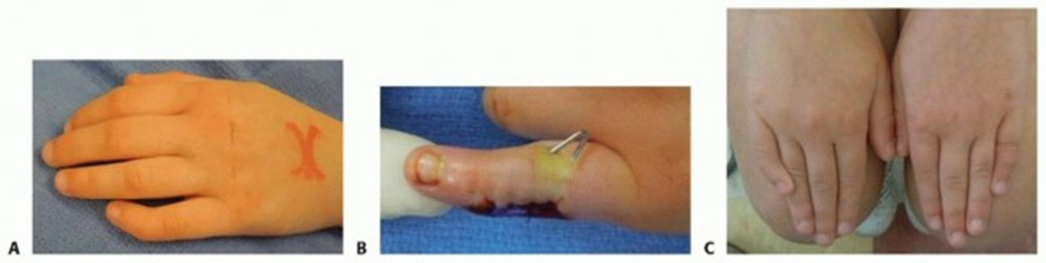
TECH FIG 2 • Small finger clinodactyly. A. Preoperative clinical photograph. B. Intraoperative photograph after osteotomy and pinning. C. Postoperative photograph demonstrating surgical correction of the left and no correction on the right.The reverse wedge is useful when large amounts of angulation correction are necessary. This technique allows for correction of angulation with preservation of length.This osteotomy is performed with an oscillating saw.A wedge of bone is taken from the near side and flipped and inserted in the far side. The osteotomy is stabilized with one or two K-wires.Undercorrection or overcorrection ofangular deformity1. Precise surgical planning with good radiographs andmeasured correctionRedundant skin causing unevenappearance of finger after phalanx excision1. Elliptical skin incisions can reduce redundant skin onthe convex side when excising a phalanx.Lack of motion after angular correction1. Excessive periosteal stripping leading to tendonadhesions. Limit soft tissue dissection.PEARLS AND PITFALLS
POSTOPERATIVE CARE
Occupational therapy is started after the first postoperative visit. The parents are instructed to wash and clean the hand. A progressive active and passive range of motion program is initiated.In cases where an osteotomy is performed, the patient is placed in a cast until the osteotomy has healed (typically 4 weeks). At this time, the pins are removed and occupational therapy is initiated.Patients are followed until full range of motion has been achieved, typically 6 to 8 weeks.
OUTCOMES
Outcomes from clinodactyly correction are generally good.Patient satisfaction is correlated to the degree of preoperative angulation and degree of correction.
COMPLICATIONS
Residual angulation may persist, usually due to initial undercorrection or continued abnormal growth. This usually is not an issue especially when the amount of angulation is mild and when the magnitude of the correction is great.Digital stiffness may be encountered. Tendon adhesions and scar tissue are usually the cause. Occupational therapy and parental education are helpful to address a loss of full digital motion.SUGGESTED READINGS1. Ali M, Jackson T, Rayan GM. Closing wedge osteotomy of abnormal middle phalanx for clinodactyly. J Hand Surg Am 2009;34: 914-918.2. Al-Qattan MM. Congenital sporadic clinodactyly of the index finger. Ann Plast Surg 2007;59:682-687.3. Bednar MS, Bindra RR, Light TR. Epiphyseal bar resection and fat interposition for clinodactyly. J HandSurg Am 2010;35:834-837.4. Strauss NL, Goldfarb CA. Surgical correction of clinodactyly: two straightforward techniques. Tech Hand Up Extrem Surg 2010;14: 54-57.5. Ty JM, James MA. Failure of differentiation: part II (arthrogryposis, camptodactyly, clinodactyly, Madelung deformity, trigger finger, and trigger thumb). Hand Clin 2009;25:195-213.
