Chapter 28Distal Femoral Physeal Fractures Martin J. Herman
DEFINITION
Fractures of the distal femoral physis are those that involve the physis or growth plate of the distal femur. These fractures occur most commonly in older children and adolescents from falls or sports activities.
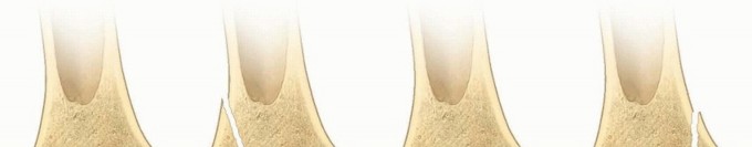
Physeal fractures of the distal femur are best categorized by the Salter-Harris (SH) classification. The vast majority of these fractures are SH type I and II fractures, which are extra-articular. SH III and IV
fractures, which are uncommon, are intra-articular (
FIG 1
).3
The goals of treatment for these fractures are healing of the fracture in acceptable alignment and anatomic restoration of the physis and joint line to reduce the risk of growth arrest and premature arthritis of the knee.
ANATOMY
The distal femoral physis accounts for 40% of the longitudinal growth of the lower extremity, growing approximately 9 mm per year until skeletal maturity.
Morphologically, this growth plate is not flat but instead has undulations across its surface which add to the stability of the physis but also make it more prone to damage when fractures of the physis occur.
The medial and lateral collateral ligaments originate from the distal femoral epiphysis distal to the physis. The anterior and posterior cruciate ligaments originate from the intercondylar notch, also distal to the physis (
FIG 2
).
The popliteal artery courses along the posterior surface of the distal femur as its traverses the popliteal space. The sciatic nerve divides into the peroneal and posterior tibial branches just proximal to the physis. Anterior displacement of the distal fragment is associated with popliteal artery injury, and medial displacement is associated with peroneal nerve injury.


FIG 1 • Patterns of distal femoral physeal fractures based on the SH classification.
PATHOGENESIS
Physeal fractures generally cleave through the zone of hypertrophic calcification then go either proximal (SH I and II) or distal to this zone (SH III and IV). In distal femoral physeal fractures, however, the fracture cleaves not only through the hypertrophic zone but also crosses other zones including the germinal zone of the growth plate because of its undulating morphology, making growth disturbance likely even after SH I and II fractures.
These fractures result most commonly from medial or lateral forces applied to the knee, resulting in varus (medial) or valgus (lateral) displacement, respectively.
Knee hyperextension injuries lead to anteriorly displaced fractures while direct forces applied to the flexed knee, such as a dashboard strike during a motor vehicle crash, commonly cause intra-articular SH III and IV fractures.
NATURAL HISTORY
The distal femoral physis has tremendous healing and remodeling potential in children with at least 2 years of growth remaining. In this age group, fractures with anatomic realignment of the joint surface that are realigned with less than 10 degrees of deformity in the anteroposterior (AP) and lateral planes heal with restoration of normal function in most patients who do
not
develop a growth arrest.
For patients who develop a growth arrest, however, the results are variable. The growth disturbance is the result of either injury to the germinal cells of the physis from the trauma of the initial injury or subsequent reduction, from malreduction with physeal bar formation, or from iatrogenic injury from screws that cross the
physis.1, 2, 6
The resulting problems related to growth disturbance are angular deformities from incomplete arrest or limb length discrepancy from complete physeal closure.


FIG 2 • AP (A) and lateral (B) diagrams of the ligaments of the knee. The collateral and cruciate ligaments have their origins distal to the distal femoral physis. C. In a child with an open physis, laxity with valgus stress testing occurs through a physeal fracture more commonly than through a medial collateral ligament tear.
Because of the high risk of growth-related problems, patients and their families must be counseled at the time of initial treatment about the potential for complications.
PATIENT HISTORY AND PHYSICAL EXAMINATION
A complete history includes an inquiry about the precise mechanism of injury and when it occurred, questions about changes in motor or sensory function of the affected limb, and any significant medical history.
A complete physical examination begins with inspection of the limb for deformity, swelling, knee joint effusion, excessive bleeding from an open injury, and any other defects of the soft tissue such as lacerations or abrasions. The entire limb is then palpated to identify focal areas of tenderness about the knee and any associated injuries proximal and distal to it.
A motor and sensory examination is necessary to identify neurologic deficits of the peroneal and posterior tibial nerves. The vascular status of the limb is assessed by assessing the distal pulses and other signs of limb perfusion including the capillary refill of the toes and the temperature of the limb.
In patients without obvious deformity but with a history and examination that is otherwise suspicious for a distal physeal fracture, gentle varus-valgus and anterior drawer or Lach-man stress testing may allow the examiner to differentiate between a physeal injury and a ligament injury.
IMAGING
High-quality AP and lateral radiographs of the entire extremity are necessary to fully assess these injuries as well as the overall alignment of the limb and other associated fractures (
FIG 3
). Dedicated views of the knee, with comparison views of the uninjured side if necessary, are useful to precisely define the fracture pattern, especially when the fracture is nondisplaced.
Computed tomography (CT) of the knee is indicated for most intra-articular fractures (SH types III and IV) to define the fracture pattern and the degree of displacement as well as to aid in planning of fixation (
FIG 4
).5
Magnetic resonance imaging (MRI) is used to confirm occult fractures when the radiographs are normal but the examination is suspicious for a fracture as well as to diagnose other knee pathology such as meniscus
tears, ligament tears, and osteochondral injuries.4
DIFFERENTIAL DIAGNOSIS
Distal femoral metaphyseal fracture Knee (tibiofemoral) dislocation Patellar dislocation
Proximal tibial fracture
Collateral ligament tears (vs. nondisplaced fracture)
NONOPERATIVE TREATMENT
Long-leg cast immobilization for 4 to 6 weeks is indicated only for fractures which are nondisplaced.
Fractures which require reduction to achieve satisfactory alignment are generally unstable and are best treated with fixation.
SURGICAL MANAGEMENT
Indications
Surgical reduction and fixation are indicated for all displaced distal femoral physeal fractures.
Surgical fixation is indicated for some nondisplaced fractures that are at high risk for displacement, such as SH III and IV fractures or those that are associated with severe soft tissue injury or neurovascular abnormalities. Other indications are obesity that inhibits effective long-leg cast immobilization and behavioral or intellectual problems that preclude cooperation with non-weight-bearing instructions.


FIG 3 • A,B. AP and lateral radiographs of displaced SH I distal femoral physeal fracture. C,D. AP and lateral radiographs of displaced SH II distal femoral physeal fracture. E,F. AP and lateral radiographs of a displaced SH III distal femoral physeal fracture.
Preoperative Planning
A vascular consultation is best called prior to going to the operating room for those patients with diminished pulses or no limb perfusion, so no intraoperative delay occurs if blood flow does not return after reduction and fixation.
The surgeon should request muscle relaxation from the anesthesia provider after induction to facilitate reduction.
The implants necessary to perform the operation include cannulated screws (4.5 to 7.3 mm in diameter),
smooth wires (5/64 inch and larger in diameter), and, in rare cases, a distal femoral plating system. Other essential equipment includes instruments to perform an open reduction and a traction bow if the need arises to place a proximal tibial traction pin for achieving length.

FIG 4 • CT images (A, coronal; B, sagittal; C, axial) of a displaced SH III fracture.
Patient Positioning
We typically perform the procedure supine on a radiolucent operating table that permits AP and lateral fluoroscopic views with the leg held in extension or with the knee flexed over a bolster or bump (
FIG 5
).
Alternatively, the patient may be positioned on a fracture table with the affected limb placed in a traction boot.


FIG 5 • Views of side (A) and foot (B) of operating table showing the patient's knee flexed over a bump and the C-arm overhead.
Surgical Approach
SH I and II Fractures
Most fractures can be realigned by closed reduction alone. The reduction maneuver entails primarily application of traction to disimpact the physis from the metaphysis and to prevent “scraping” of the two fragments together during reduction, which may exacerbate the physeal damage.
After traction is applied, appropriate translational forces are then used to achieve a gentle closed reduction. Varus or valgus forces applied to the knee reduce valgus and varus deformities, respectively. Anterior displacement is reduced by flexing the knee, whereas posterior displacement, an uncommon direction of deformity, is reduced by knee extension.
Crossed transphyseal smooth wires placed either retrograde or antegrade are best for stabilizing SH I fractures and those SH II fractures with a small Thurston-Holland fragment. Transverse metaphyseal screws that capture larger Thurston-Holland fragment are best for stabilizing SH II fractures.
Open reduction, if necessary, is performed through a longitudinal incision made at the apex of the deformity, typically at the distal end of the proximal fragment. Care must be taken to minimize physeal injury. Entrapped periosteum and soft tissue are impediments to reduction.
SH III and IV Fractures
Minimally displaced fractures may be reduced with a large reduction forceps or a stout wire used as a “joystick.”
Open reduction performed via an arthrotomy can be done either medially or laterally depending on the location of fracture extension into the joint. Anatomic realignment of the physis and the joint line are the goals of open reduction.
Stable fixation with epiphyseal screws or transphyseal smooth wires is used to stabilize these fractures.
TECHNIQUES
-
Closed Reduction and Percutaneous Wire Fixation
Fracture Reduction
Reduction is done emergently for neurovascular compromise, skin tenting, and open fractures. Otherwise, the reduction may be done the next day but no longer than 7 to 10 days after injury.
Anesthesia with muscle relaxation makes reduction easier and less traumatic to the physis.
Laterally displaced fractures are reduced by applying traction, then a medially directed force on the distal tibia while stabilizing the limb with a lateral force at the distal femur (
TECH FIG 1A
).
Anteriorly displaced fractures are reduced by applying traction to disimpact the physis and flexion of the knee (
TECH FIG 1B
).
The opposite forces are used for posterior and medial fracture displacement.
Fixation
Smooth Kirschner wires (larger than 2 mm in diameter) are used for fixation of SH I fractures and SH II fractures with small Thurston-Holland fragments. A crossed-pin configuration is commonly employed.
Wires may be placed retrograde, starting in the epiphysis and advanced across the physis into the proximal metaphyseal cortices, or antegrade, starting in the proximal metaphyseal cortices and advanced
across the physis into the epiphysis.
Wires left protruding from the joint are at high risk for causing septic arthritis of the knee. Wires should either be cut short and buried beneath the skin within the joint or advanced proximally until the wire ends are proximal to the joint cartilage but fixed in the epiphysis, making it possible to bend and cut them outside the skin adjacent to the metaphyses (
TECH FIG 2
). Buried wires are removed in the operating room, whereas percutaneous wires are removed in the office.
After fixation, the limb is immobilized in a long-leg bent knee cast or splint.


TECH FIG 1 • Coronal (A) and sagittal (B) views of an anteriorly and laterally displaced SH II fractures and the forces necessary to achieve reduction.


TECH FIG 2 • AP diagrams. A. Wire starting in medial femoral condyle drilled retrograde. B. Wire starting in medial femoral condyle drilled retrograde across proximal contralateral metaphysis and out skin. C. Wire drilled retrograde from proximal until distal end of wire is buried in the epiphysis. D. Wire starting in lateral femoral condyle drilled retrograde across proximal contralateral metaphysis and out skin. E. Drilled retrograde across proximal contralateral metaphysis and out skin. F. Wire cut and left outside the skin proximally.
-
Closed Reduction and Percutaneous Screw Fixation
Cannulated screws that do not cross the physis are most commonly used to stabilize SH II fractures with a large Thurston-Holland fragment and SH III and IV fractures.
Alternatively, smooth wires can also be used and can be placed across the physis if necessary.
Although closed reduction and percutaneous fixation is used routinely for SH II fractures, only SH III and IV fractures with minimal separation or rotation are amenable to this technique.
SH II Fractures
After closed reduction, two guidewires are placed parallel to the physis, engaging the Thurston-Holland fragment so that short-thread cannulated screws can be placed to compress across the fracture site (
TECH FIGS 3
and
4
).
Once adequate reduction and guidewire placement are confirmed with biplanar fluoroscopic views, the screws are placed sequentially, first overdrilling the outer cortex before placing the screw.
Additional fixation may be added if the fracture is unstable when stressed under fluoroscopy. The addition of a stout wire transphyseal is often my preference at this point.
Minimally Displaced SH III and IV
A large bone forceps can be used to compress across epiphyseal fragments that are separated but otherwise nondisplaced prior to guidewire placement.
For those with some rotation, the guidewire can also be used to manipulate the fragments prior to drilling the guidewire (
TECH FIG 5
).
If anatomic alignment of the physis and joint line cannot be achieved, then open reduction is required. After fixation, the limb is immobilized in a long-leg bent knee cast or splint.


TECH FIG 3 • AP diagrams. A. Displaced SH type II fracture of distal femur. B. Reduced SH type II fracture of distal femur. C. Guidewires placed across Thurston-Holland fragment, parallel to physis. D. Drill over guidewires. E. Lag screws in place.

---
TECH FIG 4
• AP (
A
) and lateral (
B
) radiographs of the patient in
FIG 3C,D
after closed reduction and fixation with 7.3-mm cannulated screws.

---
TECH FIG 5
• AP (
A
) and lateral (
B
) radiographs of the patient in
FIG 3E,F
after closed reduction and percutaneous screw fixation.
-
Open Reduction and Internal Fixation
SH I and II Fractures
Unacceptable closed reduction parameters vary to some degree by age of the child. For those with more than 2 years of growth remaining, less than 5 to 10 degrees of deformity in either plane with minimal
translation or gapping open of the physis is acceptable. Failure to achieve a near-anatomic closed reduction, however, may indicate periosteal interposition, a risk factor for growth arrest, and is an indication in many cases to perform open reduction. Children near skeletal maturity should be reduced anatomically.
Open reduction is performed under tourniquet. The incision is made either medially or laterally at the site of periosteal disruption, that is, at the apex of the deformity.
Care must be taken to remove the interposed periosteum and soft tissue (
TECH FIG 6
) and reduce the fracture without causing further damage to the physis.


TECH FIG 6 • AP diagrams of displaced SH type II fracture. A. Interposed soft tissue. B. Reduction with removal of interposed tissue.
Fixation is performed, as described earlier, based on the fracture pattern ( TECH FIG 7 ). After fixation, the limb is immobilized in a long-leg bent knee cast or splint.
SH III and IV Fractures
Open reduction is indicated for all displaced fractures that cannot be closed reduced or have complex fracture patterns.
Open reduction is performed under tourniquet. The fracture is approached through a parapatellar arthrotomy made on the side of the intra-articular fracture line.
After evacuation of the hemarthrosis, careful open reduction and fixation is performed, taking care to avoid further damage to the joint cartilage and physis.
Fixation is performed with cannulated screws placed in the epiphysis or smooth wires placed transphyseal if necessary (
TECH FIG 8
).
After fixation, the limb is immobilized in a long-leg bent knee cast or splint.

 TECH FIG 7
• AP (
A
) and lateral (
B
) radiographs of the patient in
FIG 3A,B
after open reduction and fixation with smooth wires.
TECH FIG 7
• AP (
A
) and lateral (
B
) radiographs of the patient in
FIG 3A,B
after open reduction and fixation with smooth wires.

TECH FIG 8 • AP diagrams of a displaced SH type III fracture (A) that has been lagged together (B).
PEARLS AND PITFALLS
Indications
1. Fractures that present later than 7-10 days after injury are best treated without
reduction and surgery and instead are allowed to heal. Late manipulation increases the risk of growth arrest.
Examination ▪ A careful and thorough neurovascular examination is mandatory before
manipulation to identify vascular injury or neurapraxias, especially for severely displaced fractures.
Surgical
technique
1. Fracture reduction requires longitudinal traction initially, followed by translational
maneuvers to prevent physeal damage.
2. Transphyseal screws inhibit growth and should not be used to stabilize distal femoral physeal fractures.
3. Tension on the skin around pins cut outside the skin should be relieved by extending the incision around the pin to prevent skin necrosis, which increases the risk of infection of the pins.
Follow-up
1. High-quality radiographs of the knee and a lower extremity scanogram done at 6
month intervals after injury are the best ways to identify signs of early growth disturbance.
POSTOPERATIVE CARE
After fixation, the limb is immobilized in a cast or a locked hinged knee brace for 4 to 6 weeks, and instructions are given for non-weight bearing on crutches or a walker.
Knee range of motion, strengthening exercises, and progressive weight bearing is initiated after the period of immobilization, typically under the direction of a physical therapist.
All smooth wires are removed either in the clinic within 4 to 6 weeks of surgery for those cut outside the skin or in the operating room for those buried subcutaneously. Screws are removed only for irritation or to improve the quality of imaging with CT or MRI for those with potential complications such as growth arrest and knee ligament or cartilage injuries.
OUTCOMES
For children with fractures that heal without complications and have no associated knee injuries, full return of activities and resumption of normal growth is expected.
As many as 40% to 50% of children with distal femoral physeal fractures will have a complication that requires further care or surgery to manage the complication.
COMPLICATIONS
Knee stiffness after rehabilitation is uncommon but may require prolonged therapy, manipulation, and in rare cases, knee arthroscopy and soft tissue releases.
As many as 40% of patients have associated knee injuries. Anterior cruciate ligament tear is most common and occurs most frequently after SH III and IV fractures that involve the medial condyle.
Neurovascular injuries are rare. Popliteal artery injury is associated with severely displaced fractures with anterior displacement. Peroneal nerve injury is associated with fractures that are displaced medially (varus deformity).
Growth disturbance occurs in about one-half of children with distal femoral physeal fractures.2 Careful follow-up at 4 to 6 months intervals until skeletal maturity is recommended.
ACKNOWLEDGMENT
I acknowledge the contribution of R. Dale Blasier, the author of this chapter for the first edition.
Scientific References
- 1. Arkader A, Warner WC Jr, Horn BD, et al. Predicting the outcome of physeal fractures of the distal femur. J Pediatr Orthop 2007;27:703-708 [View Source / PubMed]
- 2. Basener CJ, Mehlman CT, DiPasquale TG. Growth disturbance after distal femoral growth plate fractures in children: a meta-analysis. J Orthop Trauma 2009;23(9):663-667. [View Source / PubMed]
- 3. Beaty JH, Kumar A. Fractures about the knee in children. J Bone Joint Surg Am 1994;76(12):1870-1880. [View Source / PubMed]
- 4. Bertin KC, Goble EM. Ligament injuries associated with physeal fractures about the knee. Clin Orthop Relat Res 1983;(177): 188-195. [View Source / PubMed]
- 5. Lippert WC, Owens RF, Wall EJ. Salter-Harris type III fractures of the distal femur: plain radiographs can be deceptive. J Pediatr Orthop 2010;30(6):598-605. [View Source / PubMed]
- 6. Thomson JD, Stricker SJ, Williams MM. Fractures of the distal femoral epiphyseal plate. J Pediatr Orthop 1995;15:474-478. [View Source / PubMed]
