Introduction to Total Ankle Arthroplasty
Total Ankle Arthroplasty (TAA) represents one of the most challenging and rapidly evolving frontiers in joint replacement surgery. Historically, first-generation highly constrained and cemented ankle prostheses yielded unacceptably high rates of aseptic loosening, subsidence, and catastrophic failure. However, a paradigm shift toward unconstrained and semi-constrained cementless designs—incorporating both fixed-bearing and mobile-bearing kinematics—has revolutionized the management of end-stage ankle arthritis.
While ankle arthrodesis remains the gold standard for pain relief in the arthritic ankle, TAA offers the distinct advantage of preserving tibiotalar motion, thereby protecting adjacent hindfoot and midfoot joints from accelerated degenerative changes. This comprehensive masterclass delineates the biomechanical foundations, meticulous surgical techniques, and evidence-based outcomes associated with modern Total Ankle Arthroplasty.
Biomechanics and Implant Design
The human ankle is not a simple hinge; it is a highly complex, multi-axial joint that experiences significant shear and rotational forces during the gait cycle. The articular surface area of the talus is relatively small compared to the knee or hip, meaning the ankle joint must withstand exceptionally high contact stresses—often exceeding five times body weight during normal ambulation.
Evolution of Prosthetic Design
Modern TAA implants are broadly categorized into two distinct biomechanical philosophies:
- Fixed-Bearing Prostheses: These two-component systems (e.g., the Agility total ankle) feature a polyethylene liner locked into the tibial tray. They rely on syndesmotic fusion and a broader base of support to distribute loads.
- Mobile-Bearing Prostheses: These three-component systems (e.g., STAR, Buechel-Pappas) utilize a freely gliding polyethylene meniscus interposed between the metallic tibial and talar components. This design uncouples shear and rotational forces from the bone-implant interface, theoretically reducing the risk of aseptic loosening.
Clinical Pearl: Mobile-bearing designs require meticulous ligamentous balancing. Because the polyethylene meniscus is unconstrained, asymmetric ligamentous tension will inevitably lead to edge-loading, meniscal subluxation, or catastrophic bearing spin-out.
Below is an illustration of the Buechel-Pappas total ankle replacement device, demonstrating the three-component mobile-bearing philosophy.
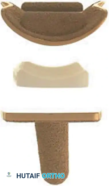
Fig. 1: Buechel-Pappas total ankle replacement device (Exploded View), highlighting the tibial tray, the mobile polyethylene meniscal bearing, and the anatomically contoured talar dome.
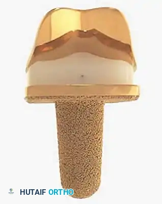
Fig. 2: Buechel-Pappas total ankle replacement device (Assembled View), demonstrating the congruent articulation designed to minimize contact stresses while permitting multi-planar motion.
Patient Selection and Indications
The success of TAA is inextricably linked to rigorous patient selection. The ideal candidate is an older, lower-demand patient with end-stage osteoarthritis, rheumatoid arthritis, or post-traumatic arthritis, who possesses good bone stock and a well-aligned hindfoot.
Primary Indications
- End-stage osteoarthritis or rheumatoid arthritis of the tibiotalar joint.
- Post-traumatic arthritis with preserved bone stock.
- Patients with symptomatic adjacent joint disease (subtalar or talonavicular arthritis) where an ankle fusion would severely compromise overall foot biomechanics.
Absolute Contraindications
- Active or recent deep infection of the ankle.
- Neuropathic arthropathy (Charcot joint).
- Avascular necrosis (AVN) involving more than 50% of the talar body.
- Severe peripheral vascular disease or compromised soft tissue envelope.
- Absent or non-functional musculature (e.g., severe paralytic deformity).
Relative Contraindications
- Severe coronal plane deformity (>15 degrees of varus or valgus) that cannot be corrected with concurrent soft tissue or bony procedures.
- Young, highly active patients or heavy manual laborers.
- Significant osteoporosis.
Preoperative Planning and Angiosome Principles
Preoperative evaluation must include weight-bearing anteroposterior (AP), lateral, and mortise radiographs of the ankle, as well as full-length standing tibial films to assess the mechanical axis. Advanced imaging (CT scan) is highly recommended to evaluate talar bone stock, subchondral cysts, and rotational profiles.
Surgical Warning: The anterior soft tissue envelope of the ankle is notoriously tenuous. Adherence to angiosome principles is critical. The anterior incision relies on the vascular territory supplied by the anterior tibial artery. Avoid undermining the skin flaps, and ensure full-thickness dissection directly down to the extensor retinaculum to preserve the subdermal vascular plexus.
Surgical Technique: The Anterior Approach
The standard anterior approach is utilized for the majority of modern TAA systems. This approach provides excellent exposure of the tibiotalar joint while allowing for precise alignment of the cutting jigs.
1. Patient Positioning and Preparation
- Place the patient supine on the operating table.
- Place a bump under the ipsilateral hip to internally rotate the leg until the patella is facing directly anteriorly. This neutralizes the natural external tibial torsion and aligns the ankle mortise.
- Apply a well-padded thigh tourniquet.
- Administer prophylactic intravenous antibiotics prior to tourniquet inflation.
2. Incision and Superficial Dissection
- Make a 10 to 15 cm longitudinal incision centered over the anterior ankle joint, lateral to the crest of the tibia and extending toward the base of the second metatarsal.
- Carry the dissection sharply through the skin and subcutaneous tissue.
- Identify and protect the superficial peroneal nerve, which typically crosses the surgical field from lateral to medial in the distal aspect of the incision.
3. Deep Dissection and Retinacular Release
- Incise the superior and inferior extensor retinaculum longitudinally.
- Develop the interval between the extensor hallucis longus (EHL) tendon medially and the extensor digitorum longus (EDL) tendons laterally.
- Retract the EHL medially and the EDL laterally.
- Identify the neurovascular bundle (anterior tibial artery and deep peroneal nerve) lying just deep to the EHL. Carefully mobilize and retract the bundle laterally with the EDL to protect it throughout the procedure.
4. Joint Exposure and Osteophyte Resection
- Perform an anterior capsulotomy to expose the tibiotalar joint.
- Aggressively resect all anterior tibial and talar osteophytes. This step is crucial; retained osteophytes will obscure the true joint line, impede the seating of cutting blocks, and restrict postoperative dorsiflexion.
5. Bony Resection and Alignment
- Tibial Preparation: Attach the extramedullary alignment guide to the tibia. The cutting block must be aligned parallel to the mechanical axis of the tibia in the coronal plane and set to the implant-specific slope in the sagittal plane. Pin the block and perform the distal tibial resection.
- Talar Preparation: Depending on the implant system, talar preparation may involve a single flat cut or a series of chamfer cuts. Ensure the talar component is perfectly aligned with the longitudinal axis of the second ray to prevent rotational malalignment.
Pitfall: Over-resection of the talus is a common error that can lead to catastrophic subsidence. Resect only enough bone to accommodate the implant, preserving the dense subchondral bone plate whenever possible.
6. Soft Tissue Balancing
- Insert trial components. Assess the ankle through a full range of motion.
- If a coronal plane deformity persists, perform sequential soft tissue releases. For varus deformities, a medial deltoid release or lateral ligament reconstruction may be necessary. For valgus deformities, consider a medializing calcaneal osteotomy or medial ligamentous plication.
7. Final Implantation
- Once optimal sizing, alignment, and kinematics are confirmed with the trials, thoroughly irrigate the joint and dry the bone surfaces.
- Impact the definitive cementless tibial and talar components. Ensure rigid press-fit fixation.
- Insert the appropriate thickness polyethylene bearing.
- Confirm final stability and range of motion.
8. Closure
- Achieve meticulous hemostasis after tourniquet deflation.
- Close the extensor retinaculum over a suction drain (if preferred) to prevent bowstringing of the tendons.
- Close the subcutaneous tissue and skin with non-absorbable sutures using a tension-free technique (e.g., Allgöwer-Donati stitches).
Postoperative Protocol
The postoperative rehabilitation protocol must balance the need for soft tissue healing with the prevention of joint stiffness.
- Weeks 0-2: The patient is placed in a bulky, well-padded short leg splint in neutral dorsiflexion. Strict non-weight-bearing (NWB) status is maintained. Elevation is critical to minimize edema and protect the incision.
- Weeks 2-4: Sutures are removed at 14-21 days once the incision is fully healed. The patient is transitioned to a removable CAM boot. Gentle active range of motion (ROM) exercises (dorsiflexion and plantarflexion) are initiated. NWB status continues.
- Weeks 4-6: Progressive partial weight-bearing in the CAM boot is initiated, advancing to full weight-bearing as tolerated.
- Weeks 6-12: The patient is weaned from the CAM boot to a supportive shoe. Formal physical therapy focuses on gait training, proprioception, and maximizing ROM.
Complications and Salvage Procedures
Despite advancements in implant design, TAA is associated with a steep learning curve and a unique set of perioperative and long-term complications.
Intraoperative Complications
- Malleolar Fractures: The medial and lateral malleoli are highly susceptible to iatrogenic fracture during bone resection or implant impaction. If a fracture occurs, it must be recognized immediately and stabilized with internal fixation (screws or tension band wiring) to prevent postoperative instability.
- Neurovascular Injury: Injury to the deep peroneal nerve or anterior tibial artery can occur during deep dissection or retractor placement.
Postoperative Complications
- Wound Healing Issues: Marginal necrosis or deep infection can be devastating. Early aggressive intervention, including debridement and targeted antibiotic therapy, is mandatory.
- Aseptic Loosening and Subsidence: The most common cause of late failure. Subsidence of the talar component is frequently observed in patients with compromised bone stock or unrecognized AVN.
- Edge Loading and Bearing Wear: In mobile-bearing designs, asymmetric ligamentous tension can cause the polyethylene meniscus to subluxate or wear rapidly, necessitating revision.
Salvage Options
When a TAA fails, salvage options are complex. They include:
1. Revision TAA: Utilizing custom or stemmed revision components with structural bone grafting.
2. Arthrodesis: Conversion to a tibiotalar or tibiotalocalcaneal (TTC) fusion using a retrograde intramedullary nail and massive structural allograft (e.g., femoral head) to restore limb length.
3. Amputation: In cases of intractable deep infection or massive un-reconstructible bone loss, a below-knee amputation may be the most functional outcome for the patient.
Conclusion
Overall, improvement in surgical technique and implant design has led to markedly better clinical outcomes compared with first-generation total ankle arthroplasty. Modern cementless, anatomically contoured, and mobile-bearing prostheses have significantly reduced the incidence of early aseptic loosening. However, it is imperative for the orthopedic surgeon to recognize that TAA results do not yet compare favorably with the exceptional long-term survivorship seen in total hip and total knee arthroplasty.
The procedure demands a profound understanding of ankle biomechanics, meticulous soft-tissue handling, and precise bony preparation. While short- to mid-term results are highly encouraging—offering excellent pain relief and preservation of gait mechanics—long-term outcomes and the ultimate survivorship of third-generation implants are yet to be definitively determined. Continued registry data and prospective randomized trials will be essential in refining patient indications and optimizing the future of ankle joint replacement.
References
- Anderson T, Montgomery F, Carlsson A: Uncemented STAR total ankle prostheses, J Bone Joint Surg 84A:1321, 2003.
- Attinger CE, Cooper P, Bloom P, et al: The safest surgical incisions and amputations applying the angiosome principles and using the Doppler to assess the arterial-arterial connections of the foot and ankle, Foot Ankle Clin N Am 6:745, 2001.
- Buechel FF, Buechel FF, Pappas MJ: Eighteen-year evaluation of cementless meniscal bearing total ankle replacements, Instr Course Lect 51:143, 2002.
- Buechel FF, Buechel FF, Pappas MJ: Ten-year evaluation of cementless Buechel-Pappas meniscal bearing total ankle replacement, Foot Ankle Int 24:462, 2003.
- Clare MP, Sanders RW, Walling AK: Total ankle arthroplasty. In Orthopaedic knowledge online, AAOS, 2003.
- Conti SF, Wong YS: Complications of total ankle replacement, Clin Orthop Relat Res 391:105, 2001.
- Easely ME, Vertullo CJ, Urban WC, et al: Total ankle arthroplasty, J Am Acad Orthop Surg 10:157, 2002.
- Gill LH: Principles of joint arthroplasty as applied to the ankle, Instr Course Lect 51:117, 2002.
- Gill LH: Challenges in total ankle arthroplasty, Foot Ankle Int 25:195, 2004.
- Haskell A, Mann RA: Ankle arthroplasty with preoperative coronal plane deformity: short-term results, Clin Orthop Relat Res 424:98, 2004.
- Knecht SI, Estin M, Callaghan JJ: The Agility total ankle arthroplasty: seven to sixteen-year follow-up, J Bone Joint Surg 86A:1161, 2004.
- Kofoed H: Cylindrical cemented ankle arthroplasty: a prospective series with long-term follow-up, Foot Ankle Int 16:474, 1995.
- Kofoed H, Lundberg-Jensen A: Ankle arthroplasty in patients younger and older than 50 years: a prospective series with long-term follow-up, Foot Ankle Int 20:501, 1999.
- Kofoed H, Sorensen TS: Ankle arthroplasty for rheumatoid arthritis and osteoarthritis: prospective long-term study of cemented replacements, J Bone Joint Surg 80B:328, 1998.
- Kopp FJ, Patel MM, Deland JT, et al: Total ankle arthroplasty with the Agility prosthesis: clinical and radiographic evaluation, Foot Ankle Int 27:97, 2006.
- McGarvey WC, Clanton TO, Lunz D: Malleolar fracture after total ankle arthroplasty: a comparison of two designs, Clin Orthop Relat Res 424:104, 2004.
- Myerson MS, Miller SD: Salvage after complications of total ankle arthroplasty, Foot Ankle Clin N Am 7:191, 2002.
- Myerson MS, Mroczek K: Perioperative complications of total ankle arthroplasty, Foot Ankle Int 24:17, 2003.
- Pyevich MT, Saltzman CL, Callaghan JJ, et al: Total ankle arthroplasty: a unique design, two to twelve-year follow-up, J Bone Joint Surg 80A:1410, 1998.
- Saltzman CL, Amendola A, Anderson R, et al: Surgeon training and complications in total ankle arthroplasty, Foot Ankle Int 24:514, 2003.
- Spirt AA, Assal M, Hansen ST: Complications and failure after total ankle arthroplasty, J Bone Joint Surg 86A:1172, 2004.
- Wood PLR: Experience with the STAR ankle arthroplasty at Wrightington Hospital, UK, Foot Ankle Clin N Am 7:755, 2002.
- Wood PLR, Deakin S: Total ankle replacement: the results in 200 ankles, J Bone Joint Surg 85B:334, 2003.
- Zerahn B, Kofoed H: Bone mineral density, gait analysis, and patient satisfaction, before and after ankle arthroplasty, Foot Ankle Int 25:208, 2004.
📚 Medical References
- total ankle arthroplasty, Clin Orthop Relat Res 268:37, 1991.
- Kitaoka HB, Anderson PJ, Morrey BF: Revision of ankle arthrodesis with external fi xation for nonunion, J Bone Joint Surg 74A:1191, 1992.
- Kitaoka HB, Romness DW: Arthrodesis for failed ankle arthroplasty, J Arthroplasty 7:277, 1992.
- Lionberger DR, Bishop JO, Tullos HS: The modifi ed Blair fusion, Foot Ankle 3:60, 1982.
- Lynch AF, Bourne RB, Rorabeck CH: The long-term results of ankle arthrodesis, J Bone Joint Surg 70B:113, 1988.
- Malarkey RF, Binski JC: Ankle arthrodesis with the Calandruccio frame and bimalleolar onlay grafting, Clin Orthop Relat Res 268:44, 1991.
- Mann RA, Van Manen JW, Wapner K, et al: Ankle fusion, Clin Orthop Relat Res 268:49, 1991.
- Marcus RE, Balourdas GM, Heiple KG: Ankle arthrodesis by chevron fusion with internal fi xation and
