A 68-year-old, active male presents to the emergency department after being struck by a motor vehicle while riding his bicycle. Radiographic assessment reveals weight-bearing dome impaction as demonstrated in Figure 6–32.
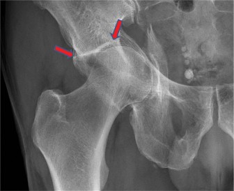
Figure 6–32
What is the ideal treatment for this patient?
- Extensile approach (iliofemoral) for ORIF of both columns
- ORIF of both columns with acute total hip arthroplasty
- Kocher-Langenbeck approach for direct reduction and fixation of the posterior column and percutaneous anterior column fixation
- Nonoperative management with skeletal traction
Discussion
The correct answer is (B). The patient’s radiographs reveal significant superior medial impaction of the acetabulum associated with this patient’s acetabular fracture. This “gull-sign” was originally described in the 60s as a radiographic representation of severe medial joint impaction. Anglen et al. evaluated this geriatric population with these specific radiographic findings and determined overall outcomes were poor despite open reduction and internal fixation. In this setting (patient >65 with significant medial joint impaction) acute total hip arthroplasty should be considered after open reduction and fixation of the acetabular fracture.
Which radiographic view is optimal for evaluation of the superomedial joint impaction that is characteristic of a geriatric acetabular fracture?
- Inlet view
- Iliac oblique view
- False profile view
- Obturator oblique view
Discussion
The correct answer is (D). The obturator oblique view is most useful for the evaluation of superomedial joint impaction in this patient population.
Objectives: Did you learn...?
Indications for considering ORIF and acute THA for an acetabular fracture?
The optimal radiographic view to visualize superomedial joint impaction in geriatric acetabular fractures?
