Biomechanics and Treatment of Femoral Shaft Fractures
Femoral shaft fractures are high-energy injuries to the femur that can result in life-threatening injuries, such as pulmonary and cerebral injuries, as well as ipsilateral femoral neck fractures. Here's what you need to know about their diagnosis, treatment, and prognosis:
Mohammad Hutaif
(
 ORCID ID: 0009-0001-1092-5600
) via
Google Scholar
.
ORCID ID: 0009-0001-1092-5600
) via
Google Scholar
.
__Save
__Cite
__Cited by
__Related articles
__All versions
__Import into BibTeX
Summary
Femoral shaft fractures are diagnosed radiographically with radiography of the femur and the hip to rule out coexisting femoral neck fractures. Treatment generally involves intramedullary nailing, which is associated with >95% union rates.
Epidemiology
Incidence: Common, with an incidence of 37.1 per 100,000 persons annually Demographics: Fractures are most common in younger patients and are often a result of high-speed motor vehicle accidents. Low-energy fractures are more common in the elderly and are often a result of a fall from standing. Gunshot fractures can also occur.
Etiology Mechanism: Traumatic and high-energy. Fracture patterns can include transverse, spiral, oblique, segmental, and comminuted fractures. Associated orthopedic injuries include ipsilateral femoral neck fracture (with an incidence of 2-6%), bilateral femur fractures, ipsilateral tibial shaft fractures, and ipsilateral acetabular fractures. Associated systemic injuries: These may include thoracic injuries such as pulmonary injury (which can lead to acute respiratory distress syndrome if early surgical treatment of the femur fracture is performed) and cerebral hemorrhage, subdural hemorrhage (which can be exacerbated by early surgical treatment of the femur fracture and intraoperative hypotension).
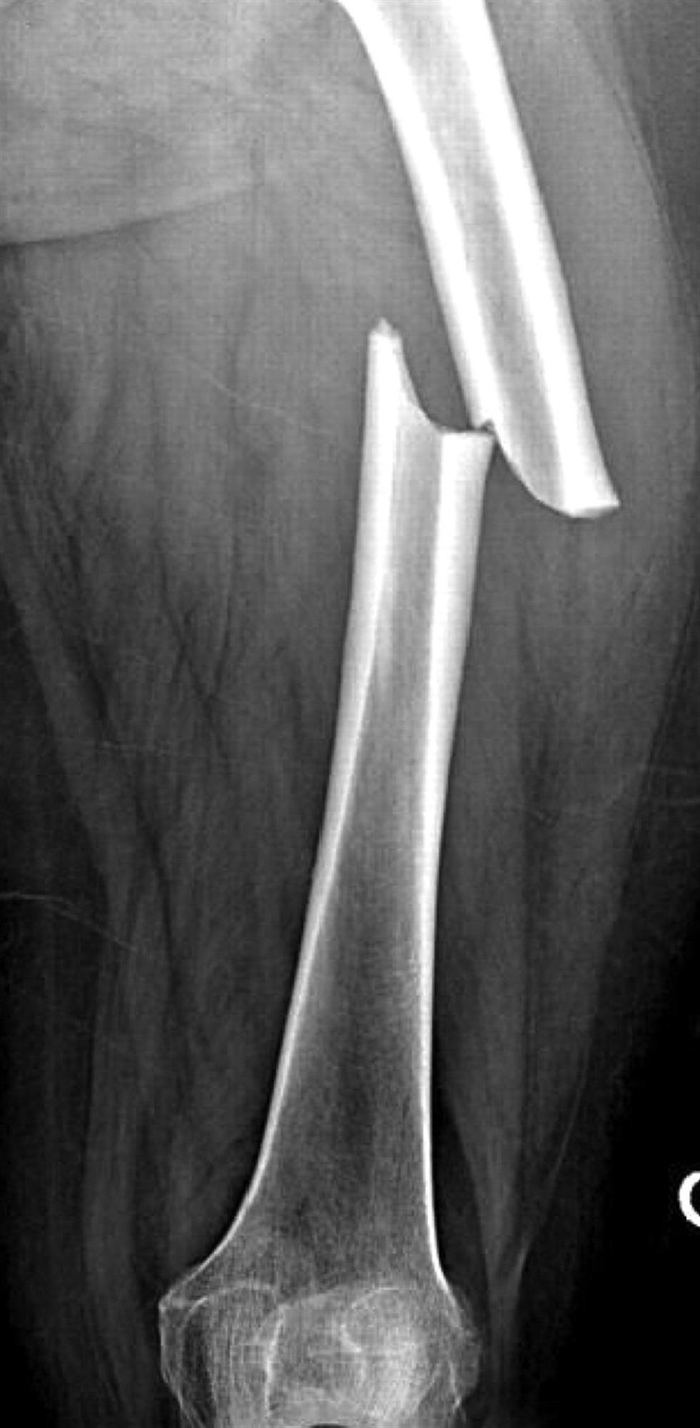
Figure 1: Biomechanics and Treatment of Femoral Shaft Fractures (Source: Hutaif Orthopedic Center)
Anatomy Osteology: The femur is the largest and strongest bone in the body. The anterior bow of the femur accommodates the musculature that surrounds it. The femoral canal runs down the length of the bone and protrudes anteriorly, forming the linea aspera. The femoral neck and subtrochanteric region are prone to fractures due to a high percentage of trabecular bone and poor blood supply. Muscles: The thigh has three compartments; the anterior, posterior, and adductor. The muscles of the compartments act as a deforming force after the fracture of the femoral shaft. The proximal fragment is abducted and flexed by the gluteus medius, minimus, and iliacus muscles. The distal fragment is subject to varus forces by the adductor muscles and extension forces from the gastrocnemius. Biomechanics: The femur is subject to pure bending, rotational, uneven bending, and four-point bending stresses depending on the fracture location and mechanism of injury.
Classification Winquist and Hansen Classification: This classification system is based on the amount of comminution, with Type 0 fractures being those with no comminution and Type IV fractures being segmental fractures with no contact between the proximal and distal fragment.
Figure 2: Winquist and Hansen Classification (Source: Hutaif Orthopedic Center)
AO/OTA Classification:
This classification system is based on the type of fracture wedge involved and is divided into three groups: Simple (32A), Wedge (32B), and Complex (32C).
Figure 3: AO/OTA Classification (Source: Hutaif Orthopedic Center)
Presentation Initial evaluation: Advanced Trauma Life Support (ATLS) should be initiated, with adequate resuscitation and normal vital signs. Initial presentation can include pain in the thigh and a tense, swollen thigh. Blood loss in a closed femoral shaft fracture can range from 1000-1500ml, and it is 500-1000ml in a closed tibial shaft fracture. On examination, there may be tenderness about the thigh, and it may be difficult to examine for an ipsilateral femoral neck fracture due to pain from the femoral shaft fracture.
Imaging Radiographs: The recommended views include AP and lateral views of the entire femur and the ipsilateral hip to rule out coexisting femoral neck fractures. CT scan may be used to rule out associated femoral neck fractures.
Treatment Nonoperative: Long leg or hip spica casting can be done for non-displaced fractures that are length-stable, or for pediatric patients with multiple medical comorbidities. Operative: Antegrade or retrograde intramedullary nailing is the gold standard for treatment. External fixation can also be used in unstable polytrauma victims or as a provisional fixation in severe open fractures.
Complications
Complications that may occur include heterotopic ossification, pudendal nerve injury, femoral artery or nerve injury, malunion and rotational malalignment, delayed union, nonunion, infection, and weakness.

Test Your Knowledge
Take the following quiz to see what you know about femoral shaft fractures: 1. What is the recommended initial evaluation for a patient with a femoral shaft fracture? MRI
CT Scan
Advanced Trauma Life Support (ATLS)
Ultrasound
2. What is the gold standard for treatment of femoral shaft fractures?
Long leg cast
Hip spica cast
Intramedullary nailing
External fixation 3. What are some of the fracture patterns seen in femoral shaft fractures? Transverse, spiral, oblique, segmental, and comminuted fractures
Buckling and stress fractures
Vertebral and sacral fractures
None of the above 4. What is the primary associated orthopedic condition seen with femoral shaft fractures? Ipsilateral acetabular fracture
Ipsilateral tibial shaft fracture
Bilateral femur fractures
Ipsilateral femoral neck fracture
5. What is the recommended radiographic evaluation for a femoral shaft fracture?
Lateral hip view
AP and lateral views of the entire femur and the ipsilateral hip
AP and lateral views of the entire femur
AP and lateral views of the ipsilateral knee 6. What is the recommended treatment for non-displaced, length-stable femoral shaft fractures in pediatric patients or patients with multiple medical comorbidities? Intramedullary nailing
Long leg cast or hip spica cast
External fixation
Open reduction internal fixation with plate 7. What are some of the complications that can occur following a femoral shaft fracture?
Heterotopic ossification, pudendal nerve injury, and femoral artery or nerve injury
Malunion and rotational malalignment, delayed union, and nonunion
Infection and weakness
All of the above
Submit
